Thymus gland
Bildnummer 11866584
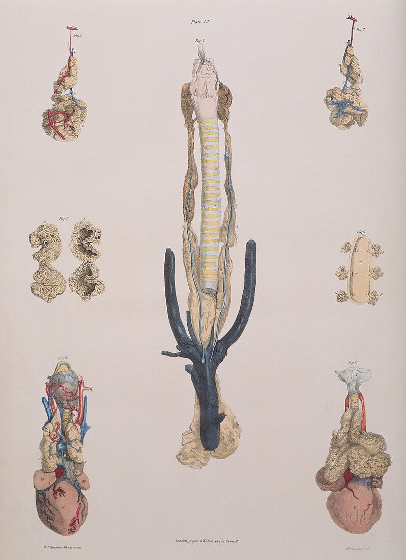
| Thymus gland. Historical illustration of the anatomy of the thymus gland. At centre is a foetal calf's thymus gland (yellow) alongside the trachea (windpipe,striped),the larynx (pink) and veins (dark blue). At upper left and right are the blood vessels (blue and red) of the thymus gland (sectioned at centre left) which is located at the base of the neck above the heart (seen at lower right and left,pink). It controls the development of lymphoid tissue in infancy and develops the white blood cells of the immune system. The internal structure is seen at centre right. Colour lithograph by Fairland from The Viscera of the Human Body,1840. From work by Sir Astley Cooper | |
| Lizenzart: | Lizenzpflichtig |
| Credit: | Science Photo Library / Terry, Sheila |
| Bildgröße: | 3206 px × 4421 px |
| Modell-Rechte: | nicht erforderlich |
| Eigentums-Rechte: | nicht erforderlich |
| Restrictions: | - |
Preise für dieses Bild ab 15 €
Universitäten & Organisationen
(Informationsmaterial Digital, Informationsmaterial Print, Lehrmaterial Digital etc.)
ab 15 €
Redaktionell
(Bücher, Bücher: Sach- und Fachliteratur, Digitale Medien (redaktionell) etc.)
ab 30 €
Werbung
(Anzeigen, Aussenwerbung, Digitale Medien, Fernsehwerbung, Karten, Werbemittel, Zeitschriften etc.)
ab 55 €
Handelsprodukte
(bedruckte Textilie, Kalender, Postkarte, Grußkarte, Verpackung etc.)
ab 75 €
Pauschalpreise
Rechtepakete für die unbeschränkte Bildnutzung in Print oder Online
ab 495 €