Ear anatomy
Bildnummer 11866521
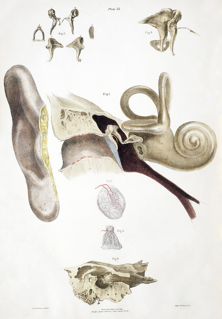
| Ear anatomy. Historical anatomical artwork of a human ear. The main diagram (centre) shows the outer ear (pinna,left) and the internal structure of the ear (moving left-right): the ear canal,the ear drum (blue),the ear bones,the cochlea (snail shaped). Just before the cochlea are two other stuctures: the semicircular canals (three loops at right angles to each other),and the Eustachian tube (brown,connecting to the throat). The ear bones are also shown at top. The ear drum and its blood vessels is also at lower centre with another ear membrane below. The temporal bone (bottom) houses the ear. Artwork from The Nerves of the Human Body (Ed. Jones Quain,London,1839) | |
| Lizenzart: | Lizenzpflichtig |
| Credit: | Science Photo Library / Terry, Sheila |
| Bildgröße: | 3283 px × 4704 px |
| Modell-Rechte: | nicht erforderlich |
| Eigentums-Rechte: | nicht erforderlich |
| Restrictions: | - |
Preise für dieses Bild ab 15 €
Universitäten & Organisationen
(Informationsmaterial Digital, Informationsmaterial Print, Lehrmaterial Digital etc.)
ab 15 €
Redaktionell
(Bücher, Bücher: Sach- und Fachliteratur, Digitale Medien (redaktionell) etc.)
ab 30 €
Werbung
(Anzeigen, Aussenwerbung, Digitale Medien, Fernsehwerbung, Karten, Werbemittel, Zeitschriften etc.)
ab 55 €
Handelsprodukte
(bedruckte Textilie, Kalender, Postkarte, Grußkarte, Verpackung etc.)
ab 75 €
Pauschalpreise
Rechtepakete für die unbeschränkte Bildnutzung in Print oder Online
ab 495 €
Keywords
- 1800er Jahre,
- 19. Jahrhundert,
- Anatomie,
- anatomisch,
- Audiologie,
- Buch,
- Geschichte,
- Hammer,
- historisch,
- Illustration,
- Jones Quain,
- Kanal,
- Knochen,
- Kunstwerk,
- Medizin,
- medizinisch,
- Mensch,
- Ohr,
- Profil,
- Schläfenbein,
- Seite,
- seitlich,
- Sektion,
- sektioniert,
- Sinn,
- Trommel,
- Trommelfell,
- Vorhalle,
- Zeichnung