Head and neck muscles
Bildnummer 11866369
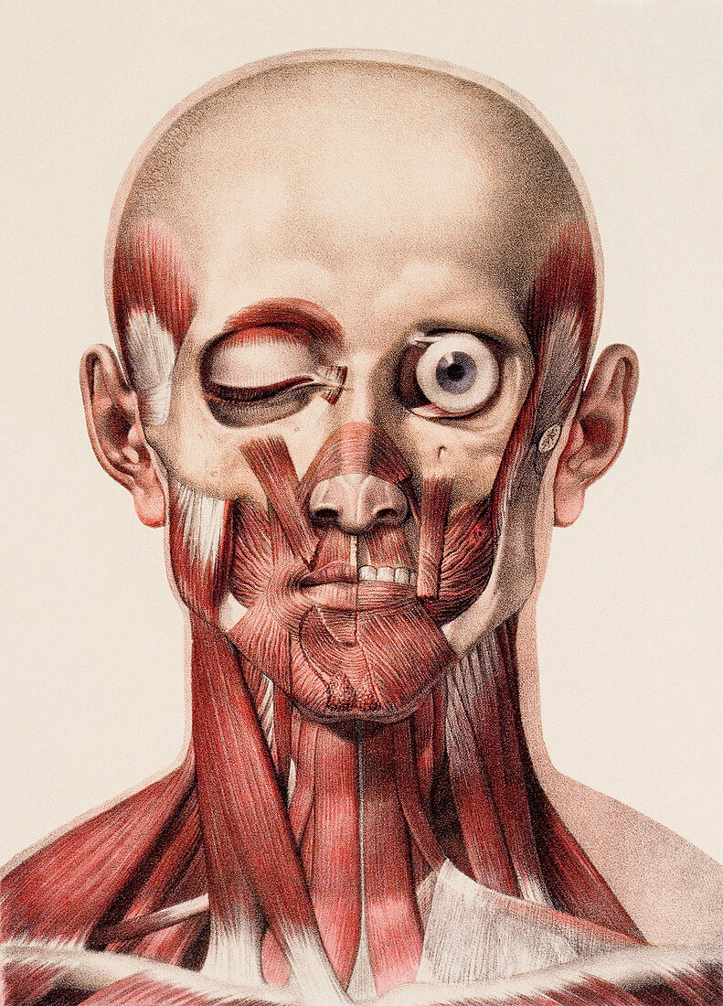
| Head and neck muscles. Historical anatomical artwork of the muscles of the human head and neck,seen from the front. The specimen has been dissected to two different levels. At left,the dissection shows the intermediate muscles of the head and the superficial muscles of the neck. At right,the deep muscle layer has been revealed after the removal of the zygomatic arch (rear part of the cheek bone) and the jaw muscle and sternomastoid neck muscle. Artwork from the 19th- century book Atlas of Anatomy,by Bourgery and Jacob. This book,which took over 20 years to complete,was published in France in 8 volumes from 1831 to 1854. It contained 726 colour plates covering both anatomy and surgical techniques | |
| Lizenzart: | Lizenzpflichtig |
| Credit: | Science Photo Library / Kulyk, Mehau |
| Bildgröße: | 3173 px × 4410 px |
| Modell-Rechte: | nicht erforderlich |
| Eigentums-Rechte: | nicht erforderlich |
| Restrictions: | - |
Preise für dieses Bild ab 15 €
Universitäten & Organisationen
(Informationsmaterial Digital, Informationsmaterial Print, Lehrmaterial Digital etc.)
ab 15 €
Redaktionell
(Bücher, Bücher: Sach- und Fachliteratur, Digitale Medien (redaktionell) etc.)
ab 30 €
Werbung
(Anzeigen, Aussenwerbung, Digitale Medien, Fernsehwerbung, Karten, Werbemittel, Zeitschriften etc.)
ab 55 €
Handelsprodukte
(bedruckte Textilie, Kalender, Postkarte, Grußkarte, Verpackung etc.)
ab 75 €
Pauschalpreise
Rechtepakete für die unbeschränkte Bildnutzung in Print oder Online
ab 495 €
Keywords
- 1800er Jahre,
- 19. Jahrhundert,
- Anatomie,
- anatomisch,
- anterior,
- aufgegliedert,
- ausgeschnitten,
- Ausschnitte,
- Biologie,
- biologisch,
- Erwachsene,
- Französisch,
- Frontal,
- Geschichte,
- Gesicht,
- Hals,
- historisch,
- Illustration,
- Jean Baptiste Marc Bourgery,
- Kopf,
- Leiche,
- Mann,
- Männer,
- Männlich,
- Medizin,
- medizinisch,
- Mensch,
- Menschen Person Personen,
- menschlicher Körper,
- Muskel,
- Muskulatur,
- Muskulös,
- Nicolas Henri Jacob,
- Zeichnung