Skull anatomy by Leonardo da Vinci
Bildnummer 11866344
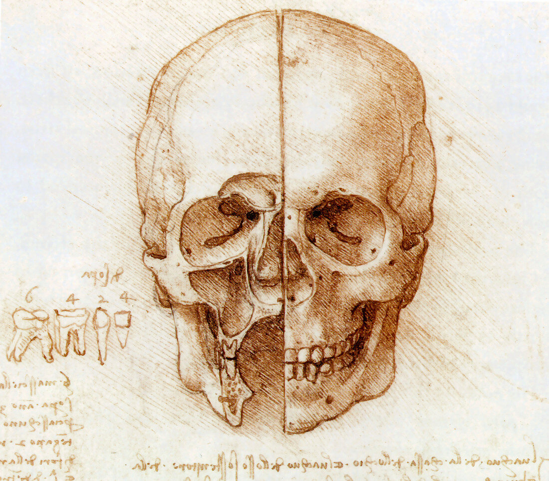
| Skull anatomy by Leonardo da Vinci. Historical artwork and notes on the anatomy of the human skull and teeth,by the Italian artist and scientist Leonardo da Vinci (1452-1519). This bisected skull shows the external structure (right),and dissected facial sinuses (left),the air-filled spaces inside the bones of the face. The diagram at lower left shows the teeth present in one half of the mouth: 4 incisors,2 canines,4 pre-molars,and 6 molars. Da Vinci was the first anatomist known to have correctly noted the number and root structure of human teeth. The notes are an example of his mirror writing,which was written backwards from right to left,and could be read in a mirror | |
| Lizenzart: | Lizenzpflichtig |
| Credit: | Science Photo Library / Terry, Sheila |
| Bildgröße: | 3168 px × 2773 px |
| Modell-Rechte: | nicht erforderlich |
| Eigentums-Rechte: | nicht erforderlich |
| Restrictions: | - |
Preise für dieses Bild ab 15 €
Universitäten & Organisationen
(Informationsmaterial Digital, Informationsmaterial Print, Lehrmaterial Digital etc.)
ab 15 €
Redaktionell
(Bücher, Bücher: Sach- und Fachliteratur, Digitale Medien (redaktionell) etc.)
ab 30 €
Werbung
(Anzeigen, Aussenwerbung, Digitale Medien, Fernsehwerbung, Karten, Werbemittel, Zeitschriften etc.)
ab 55 €
Handelsprodukte
(bedruckte Textilie, Kalender, Postkarte, Grußkarte, Verpackung etc.)
ab 75 €
Pauschalpreise
Rechtepakete für die unbeschränkte Bildnutzung in Print oder Online
ab 495 €
Keywords
- 15. Jahrhundert,
- 1500er Jahre,
- 16. Jahrhundert,
- Anatomie,
- anatomisch,
- aufgegliedert,
- Auge,
- Bleistift,
- Caninus,
- Code,
- dental,
- Diagramm,
- erst,
- europäisch,
- fazial,
- Frontal,
- genau,
- Geschichte,
- Gesicht,
- Hinweis,
- historisch,
- Hohlraum,
- Illustration,
- Italienisch,
- Jochbogen,
- Keilbein,
- Kiefer,
- Knochen,
- Kunstwerk,
- Leonardo Da Vinci,
- Mensch,
- Menschen Person Personen,
- monochromes Bild,
- Mund,
- Nasal-,
- Nase,
- Nebenhöhlen,
- Notizbuch,
- Notizbücher,
- Oral,
- Papier,
- Renaissance,
- Schädel,
- Schneidezahn,
- Schreiben,
- Schwarz und weiß,
- Siebbein,
- Sinus,
- Skelett,
- Skelett-,
- Steckdose,
- Steckdosen,
- Umlaufbahnen,
- Unterkiefer,
- Vorderseite,
- Wurzel,
- Wurzeln,
- Zahn,
- Zähne,
- Zeichnung