Removing a displaced IUD contraceptive
Bildnummer 11862998
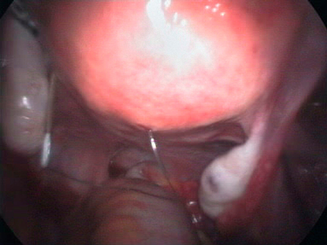
| Removal of a displaced IUD contraceptive. Endoscope view of a surgical instrument removing an intrauterine contraceptive device (IUD,white and brown,centre left) that has moved from the uterus (womb,top to centre) into the abdomen. The IUD is located in the pouch of Douglas,a part of the peritoneal cavity between the rectum and the back wall of the uterus. The ovaries (white) are seen either side of the uterus. An IUD is a T- shaped piece of metal or plastic that is inserted into the uterus,through the cervix,to prevent pregnancy. It is thought to work by inhibiting the implantation of a fertilised egg. In cases where any part of the IUD has penetrated the uterine wall,the IUD is surgically removed | |
| Lizenzart: | Lizenzpflichtig |
| Credit: | Science Photo Library / Layyous, Dr. Najeeb |
| Bildgröße: | 2910 px × 2181 px |
| Modell-Rechte: | nicht erforderlich |
| Eigentums-Rechte: | nicht erforderlich |
| Restrictions: | - |
Preise für dieses Bild ab 15 €
Universitäten & Organisationen
(Informationsmaterial Digital, Informationsmaterial Print, Lehrmaterial Digital etc.)
ab 15 €
Redaktionell
(Bücher, Bücher: Sach- und Fachliteratur, Digitale Medien (redaktionell) etc.)
ab 30 €
Werbung
(Anzeigen, Aussenwerbung, Digitale Medien, Fernsehwerbung, Karten, Werbemittel, Zeitschriften etc.)
ab 55 €
Handelsprodukte
(bedruckte Textilie, Kalender, Postkarte, Grußkarte, Verpackung etc.)
ab 75 €
Pauschalpreise
Rechtepakete für die unbeschränkte Bildnutzung in Print oder Online
ab 495 €
Keywords
- Abdomen,
- Ausrüstung,
- Bauch,
- Betrieb,
- Eierstock,
- Eierstöcke,
- Empfängnisverhütung,
- Endoskop,
- Endoskopie,
- endoskopisch,
- Entfernen,
- Erwachsene,
- Frau,
- Geburtenkontrolle,
- geduldig,
- Gerät,
- Gesundheitswesen,
- Gynäkologie,
- Instrument,
- iud,
- Kondition,
- Laparoskopie,
- Medizin,
- medizinisch,
- menschlicher Körper,
- Reproduktionspathologie,
- Spule,
- Störung,
- Verhütungsmittel,
- Weiblich,
- weibliches Fortpflanzungssystem