LM of trophoblastic cells from hydatiform mole
Bildnummer 11862716
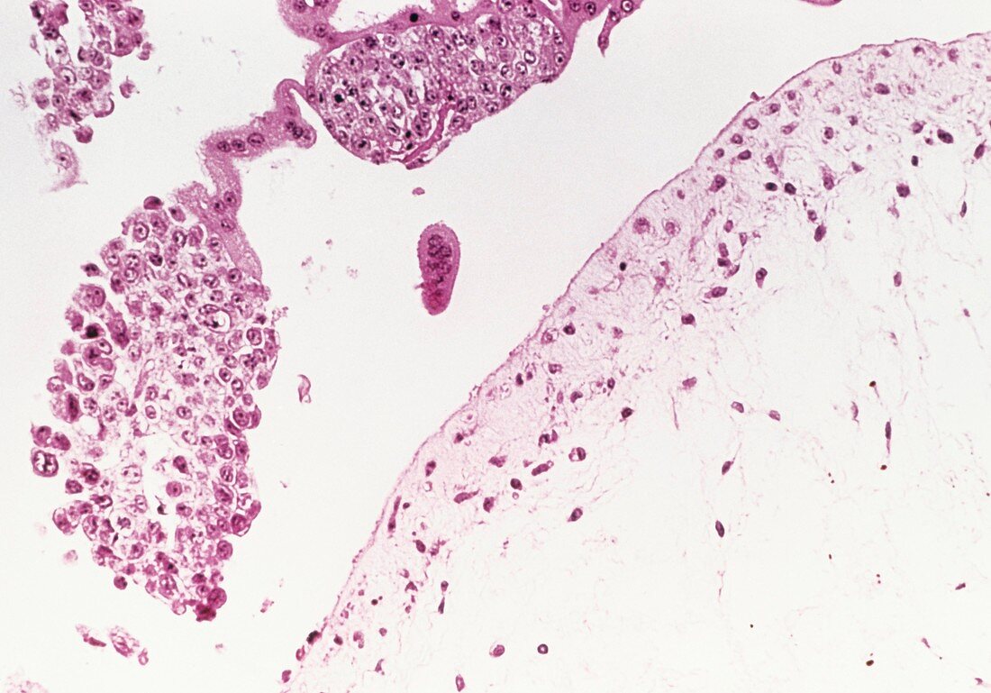
| Hydatidiform mole. Light micrograph of trophobla- stic cells from the endometrium lining of the uterus showing the formation of a hydatidiform mole. A hydatidiform mole is seen in some unsucce- ssful pregnancies. The membrane surrounding the embryo (the chorion) degenerates,forming a collection of fluid-filled sacs. Trophoblastic cells,which make up the chorion are seen here forming thick layers (at lower right) and disconnected masses (at left). The cells in the disconnected masses are of abnormal shape and size,and may go on to form a choriocarcinoma,an extremely rare and malignant tumour. Papanicolaou stained cells. Magnification: x40 at 35mm size | |
| Lizenzart: | Lizenzpflichtig |
| Credit: | Science Photo Library |
| Bildgröße: | 5020 px × 3511 px |
| Modell-Rechte: | nicht erforderlich |
| Eigentums-Rechte: | nicht erforderlich |
| Restrictions: | - |
Preise für dieses Bild ab 15 €
Universitäten & Organisationen
(Informationsmaterial Digital, Informationsmaterial Print, Lehrmaterial Digital etc.)
ab 15 €
Redaktionell
(Bücher, Bücher: Sach- und Fachliteratur, Digitale Medien (redaktionell) etc.)
ab 30 €
Werbung
(Anzeigen, Aussenwerbung, Digitale Medien, Fernsehwerbung, Karten, Werbemittel, Zeitschriften etc.)
ab 55 €
Handelsprodukte
(bedruckte Textilie, Kalender, Postkarte, Grußkarte, Verpackung etc.)
ab 75 €
Pauschalpreise
Rechtepakete für die unbeschränkte Bildnutzung in Print oder Online
ab 495 €