Coloured SEM of cells of Brenner tumour in ovary
Bildnummer 11862668
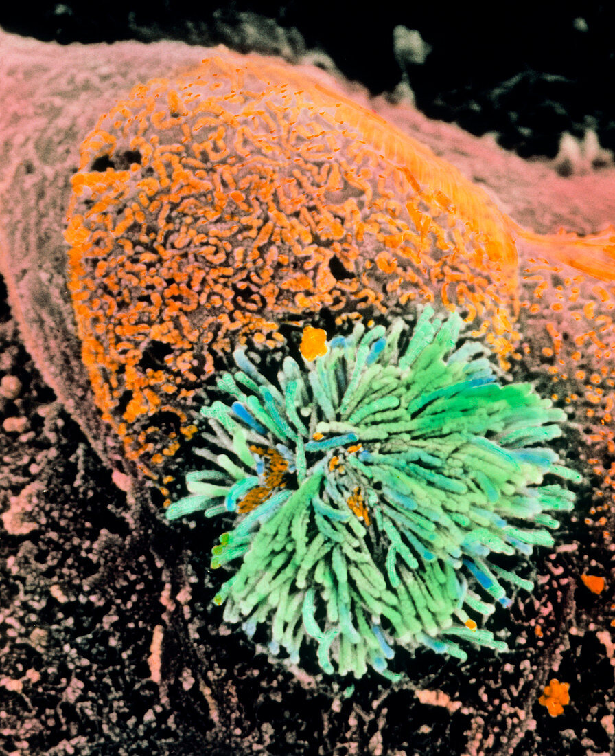
| Brenner tumour in the ovary. Coloured Scanning Electron Micrograph (SEM) of cells of a Brenner tumour on the surface of a human ovary. Tufts of cilia (green) of a ciliated cell are seen; it is located among columnar secretory cells (orange) with microvilli. These cell elements make up the Brenner tumour,a benign tumour (or fibro- epithelioma) of the surface epithelial cells of the ovary. The cells are irregularly shaped and highly proliferative. Named after the German pathologist Fritz Brenner,a Brenner tumour that is proliferative may become malignant (cancerous). Magnification: x4,060 at 6x7cm size. x5,200 at 4x5ins | |
| Lizenzart: | Lizenzpflichtig |
| Credit: | Science Photo Library / PROFESSORS P.M. MOTTA & S. MAKABE |
| Bildgröße: | 2982 px × 3659 px |
| Modell-Rechte: | nicht erforderlich |
| Eigentums-Rechte: | nicht erforderlich |
| Restrictions: | - |
Preise für dieses Bild ab 15 €
Universitäten & Organisationen
(Informationsmaterial Digital, Informationsmaterial Print, Lehrmaterial Digital etc.)
ab 15 €
Redaktionell
(Bücher, Bücher: Sach- und Fachliteratur, Digitale Medien (redaktionell) etc.)
ab 30 €
Werbung
(Anzeigen, Aussenwerbung, Digitale Medien, Fernsehwerbung, Karten, Werbemittel, Zeitschriften etc.)
ab 55 €
Handelsprodukte
(bedruckte Textilie, Kalender, Postkarte, Grußkarte, Verpackung etc.)
ab 75 €
Pauschalpreise
Rechtepakete für die unbeschränkte Bildnutzung in Print oder Online
ab 495 €