LM of biopsy section through human cervix
Bildnummer 11862586
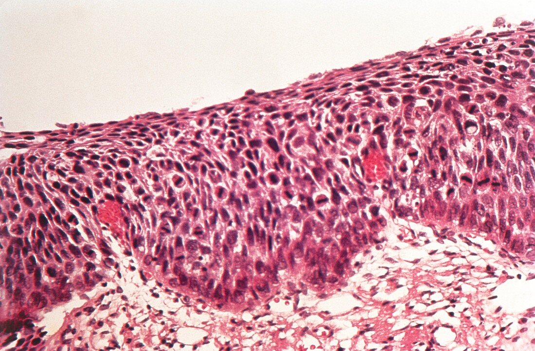
| Light micrograph of a biopsy section through the human cervix revealing carcinoma-in-situ (severe dysplasia,CIN 3). The cervix is lined by a stratified squamous epithelium. Normally,all cell division is confined to the basal epithelial layer (bottom). As a cell matures & rises up the layers,its cytoplasm expands & nucleus shrinks in size - a normal superficial cell is flat,with a small nucleus & a large cytoplasm. In carcinoma-in-situ the gradual stratification in cell maturation seen in normal epithelium is absent: dysplastic (abnormal) cells,some in process of dividing,extend throughout the entire epithelium. The basement membrane remains defined & intact | |
| Lizenzart: | Lizenzpflichtig |
| Credit: | Science Photo Library |
| Bildgröße: | 5197 px × 3411 px |
| Modell-Rechte: | nicht erforderlich |
| Eigentums-Rechte: | nicht erforderlich |
| Restrictions: | - |
Preise für dieses Bild ab 15 €
Universitäten & Organisationen
(Informationsmaterial Digital, Informationsmaterial Print, Lehrmaterial Digital etc.)
ab 15 €
Redaktionell
(Bücher, Bücher: Sach- und Fachliteratur, Digitale Medien (redaktionell) etc.)
ab 30 €
Werbung
(Anzeigen, Aussenwerbung, Digitale Medien, Fernsehwerbung, Karten, Werbemittel, Zeitschriften etc.)
ab 55 €
Handelsprodukte
(bedruckte Textilie, Kalender, Postkarte, Grußkarte, Verpackung etc.)
ab 75 €
Pauschalpreise
Rechtepakete für die unbeschränkte Bildnutzung in Print oder Online
ab 495 €