Healthy cervical smear
Bildnummer 11862578
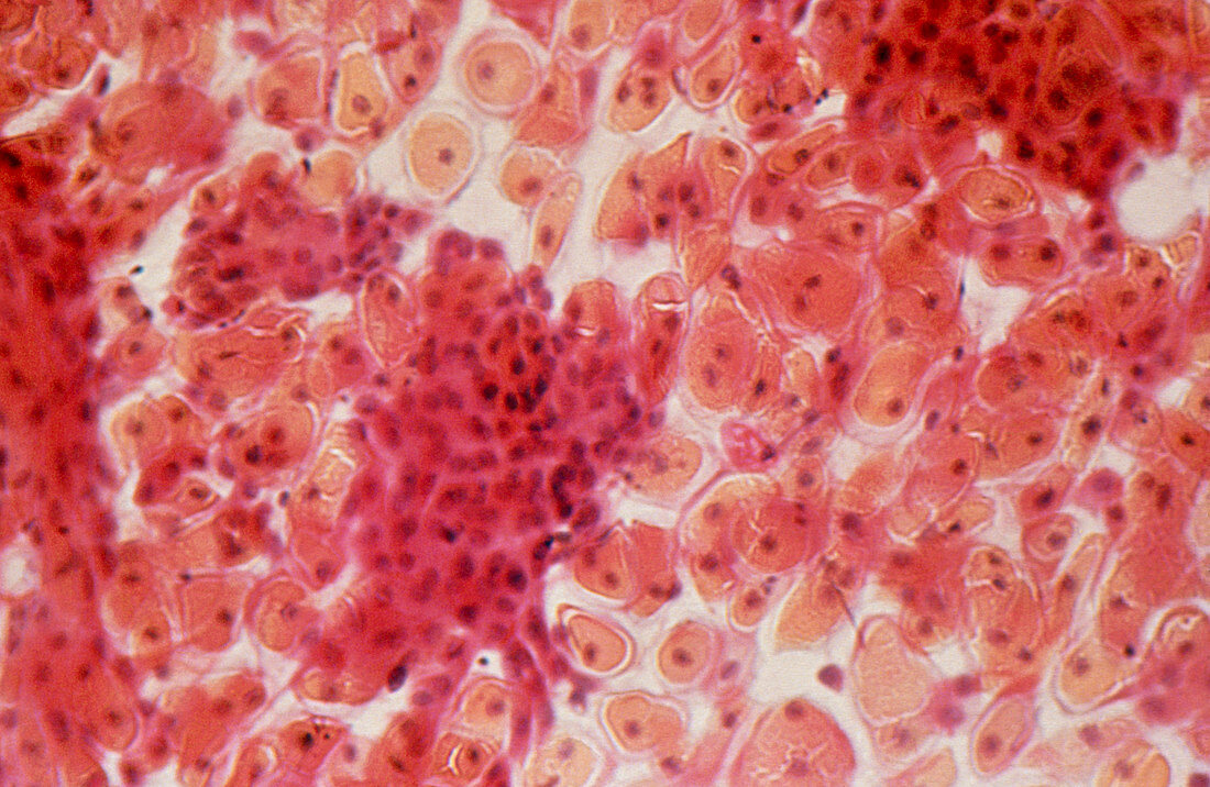
| Light micrograph of normal cells in a cervical smear taken post partum - after childbirth. The cells visible are from the intermediate and parabasal zones - the inner two of the four groups of layers that form the stratified squamous epithelium that lines the vaginal cervix. The parabasal zone consisits of several layers of large polygonal cells (orange stain). Cells of the inner intermediate zone are small & ovoid in shape. The relative thickness & proportions of these different zones varies throughout the menstrual cycle. Cervical smears are examined to alert a doctor to the cellular changes associated with the development of cervical cancer | |
| Lizenzart: | Lizenzpflichtig |
| Credit: | Science Photo Library |
| Bildgröße: | 4944 px × 3216 px |
| Modell-Rechte: | nicht erforderlich |
| Eigentums-Rechte: | nicht erforderlich |
| Restrictions: | - |
Preise für dieses Bild ab 15 €
Universitäten & Organisationen
(Informationsmaterial Digital, Informationsmaterial Print, Lehrmaterial Digital etc.)
ab 15 €
Redaktionell
(Bücher, Bücher: Sach- und Fachliteratur, Digitale Medien (redaktionell) etc.)
ab 30 €
Werbung
(Anzeigen, Aussenwerbung, Digitale Medien, Fernsehwerbung, Karten, Werbemittel, Zeitschriften etc.)
ab 55 €
Handelsprodukte
(bedruckte Textilie, Kalender, Postkarte, Grußkarte, Verpackung etc.)
ab 75 €
Pauschalpreise
Rechtepakete für die unbeschränkte Bildnutzung in Print oder Online
ab 495 €