Lm of cervical smearshowing pre-invasive cancer
Bildnummer 11862575
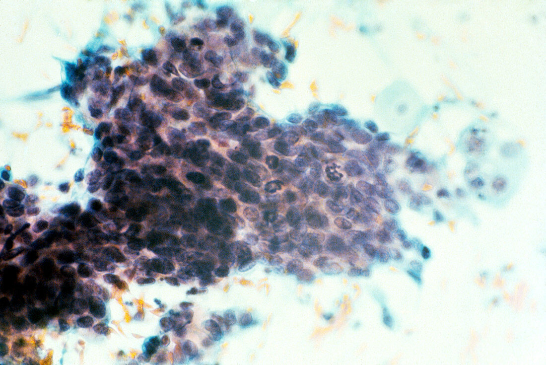
| Light micrograph from a cervical smear showing abnormal squamous epithelial cells constituting a case of carcinoma-in-situ (c.i.s.),or pre- invasive cancer of the cervix. The individual cells are severely dysplastic,being pleomorphic or of heterogenous form,with large nuclei that show evidence of cell divisions. Normally the superficial squamous cells would appear uniform with large cytoplasms and small centrally-placed nuclei,with cell division confined to the basal layer. The degrees of dysplasia range from mild,through moderate,to severe and c.i.s. They are graded CIN I,II and III respectively. Magnificat- ion: x40 at 35mm size | |
| Lizenzart: | Lizenzpflichtig |
| Credit: | Science Photo Library |
| Bildgröße: | 3711 px × 2480 px |
| Modell-Rechte: | nicht erforderlich |
| Eigentums-Rechte: | nicht erforderlich |
| Restrictions: | - |
Preise für dieses Bild ab 15 €
Universitäten & Organisationen
(Informationsmaterial Digital, Informationsmaterial Print, Lehrmaterial Digital etc.)
ab 15 €
Redaktionell
(Bücher, Bücher: Sach- und Fachliteratur, Digitale Medien (redaktionell) etc.)
ab 30 €
Werbung
(Anzeigen, Aussenwerbung, Digitale Medien, Fernsehwerbung, Karten, Werbemittel, Zeitschriften etc.)
ab 55 €
Handelsprodukte
(bedruckte Textilie, Kalender, Postkarte, Grußkarte, Verpackung etc.)
ab 75 €
Pauschalpreise
Rechtepakete für die unbeschränkte Bildnutzung in Print oder Online
ab 495 €