Knee joint prosthesis,X-ray
Bildnummer 11854157
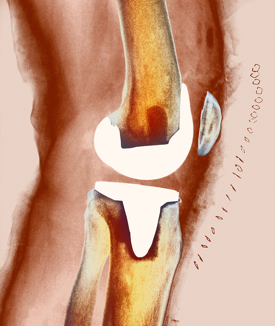
| Knee joint replacement. Coloured X-ray of the knee of a 60 year old woman (side view) showing an artificial (prosthetic) joint replacing the knee joint. The entire knee joint surface has been replaced here. The implant (white) made of metal alloy attaches to the bottom of the femur (thigh bone,upper frame) and to the top of the tibia (shin bone,lower frame). It forms a flexible joint that can hinge like the old joint,relieving joint pain and immobility. Following surgery,metal sutures (at right) are seen on the skin surface; the kneecap (patella bone) is also seen. Knee joint replacement is conducted usually because of joint disease such as osteoarthritis or due to injury | |
| Lizenzart: | Lizenzpflichtig |
| Credit: | Science Photo Library |
| Bildgröße: | 3365 px × 3988 px |
| Modell-Rechte: | nicht erforderlich |
| Eigentums-Rechte: | nicht erforderlich |
| Restrictions: | - |
Preise für dieses Bild ab 15 €
Universitäten & Organisationen
(Informationsmaterial Digital, Informationsmaterial Print, Lehrmaterial Digital etc.)
ab 15 €
Redaktionell
(Bücher, Bücher: Sach- und Fachliteratur, Digitale Medien (redaktionell) etc.)
ab 30 €
Werbung
(Anzeigen, Aussenwerbung, Digitale Medien, Fernsehwerbung, Karten, Werbemittel, Zeitschriften etc.)
ab 55 €
Handelsprodukte
(bedruckte Textilie, Kalender, Postkarte, Grußkarte, Verpackung etc.)
ab 75 €
Pauschalpreise
Rechtepakete für die unbeschränkte Bildnutzung in Print oder Online
ab 495 €
Keywords
- Arthritis,
- arthritisch,
- Arthrose,
- Behandlung,
- Bein,
- Ersatz,
- farbig,
- Femur,
- Frau,
- geduldig,
- gefärbt,
- Gesundheitswesen,
- Implantat,
- Kniescheibe,
- Knochen,
- künstliches Kniegelenk,
- Medizin,
- medizinisch,
- nach der Operation,
- Nähte,
- Prothese,
- Radiographie,
- Röntgen,
- Schenkel,
- Schienbein,
- Seitenansicht,
- Wadenbein,
- Weiblich