Hip joint replacement,X-ray
Bildnummer 11854147
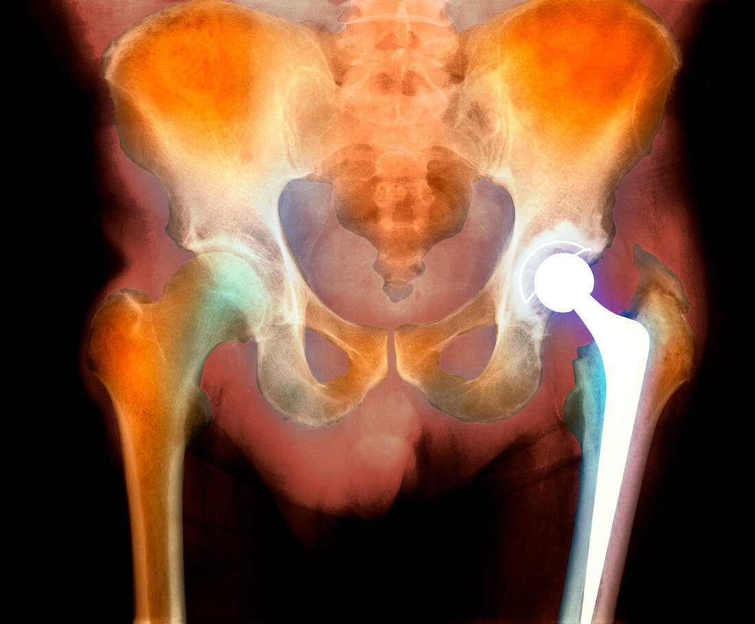
| Hip joint replacement. Coloured X-ray of the pelvis (front view) of a 73 year old man,showing an artificial or prosthetic hip joint implant (white,lower right). The hip joint is a ball-and-socket joint. The ball part of the implant is attached by a long peg into the femur bone,with surgical cement. The socket part of the joint,located in the pelvis bone,has also been replaced. The new hip joint is made of a composite metal alloy. Hip joint replacement may be needed due to disease such as arthritis,or due to an injury | |
| Lizenzart: | Lizenzpflichtig |
| Credit: | Science Photo Library |
| Bildgröße: | 4628 px × 3825 px |
| Modell-Rechte: | nicht erforderlich |
| Eigentums-Rechte: | nicht erforderlich |
| Restrictions: | - |
Preise für dieses Bild ab 15 €
Universitäten & Organisationen
(Informationsmaterial Digital, Informationsmaterial Print, Lehrmaterial Digital etc.)
ab 15 €
Redaktionell
(Bücher, Bücher: Sach- und Fachliteratur, Digitale Medien (redaktionell) etc.)
ab 30 €
Werbung
(Anzeigen, Aussenwerbung, Digitale Medien, Fernsehwerbung, Karten, Werbemittel, Zeitschriften etc.)
ab 55 €
Handelsprodukte
(bedruckte Textilie, Kalender, Postkarte, Grußkarte, Verpackung etc.)
ab 75 €
Pauschalpreise
Rechtepakete für die unbeschränkte Bildnutzung in Print oder Online
ab 495 €
Keywords
- Arthritis,
- Arthrose,
- Becken,
- Behandlung,
- Bein,
- Beine,
- Ersatz,
- farbig,
- Femur,
- geduldig,
- gefärbt,
- Gelenk,
- Gelenke,
- Gesundheitswesen,
- Hüfte,
- Hüften,
- Joint,
- Knochen,
- künstliches Implantat,
- Mann,
- Männlich,
- Medizin,
- medizinisch,
- Oberschenkelknochen,
- pelvin,
- Prothese,
- Radiographie,
- Röntgen,
- Röntgenbild,
- Vorderansicht