Knee replacement,X-rays
Bildnummer 11854125
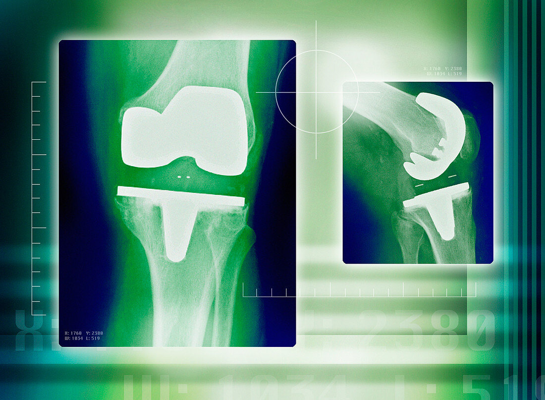
| Artificial knee. Coloured X-rays of an artificial (prosthetic) knee joint. At left is a frontal X- ray of a straight knee and at right is a side view of a bent knee. The prostheses are implanted into the thigh bone (femur) and shinbone (tibia) with pegs and surgical cement. The artificial joint hinges in an identical way to a normal knee joint. Knee joints need replacing when the protective cartilage that surrounds them is lost. Cartilage can be lost due to injury or osteoarthritis | |
| Lizenzart: | Lizenzpflichtig |
| Credit: | Science Photo Library / Maslo, Miriam |
| Bildgröße: | 4884 px × 3582 px |
| Modell-Rechte: | nicht erforderlich |
| Eigentums-Rechte: | nicht erforderlich |
| Restrictions: | - |
Preise für dieses Bild ab 15 €
Universitäten & Organisationen
(Informationsmaterial Digital, Informationsmaterial Print, Lehrmaterial Digital etc.)
ab 15 €
Redaktionell
(Bücher, Bücher: Sach- und Fachliteratur, Digitale Medien (redaktionell) etc.)
ab 30 €
Werbung
(Anzeigen, Aussenwerbung, Digitale Medien, Fernsehwerbung, Karten, Werbemittel, Zeitschriften etc.)
ab 55 €
Handelsprodukte
(bedruckte Textilie, Kalender, Postkarte, Grußkarte, Verpackung etc.)
ab 75 €
Pauschalpreise
Rechtepakete für die unbeschränkte Bildnutzung in Print oder Online
ab 495 €
Keywords
- Behandlung,
- Bein,
- Biegen,
- Diagnose,
- Duo,
- Ersatz,
- farbig,
- Femur,
- gefärbt,
- Gelenk,
- Gesundheitswesen,
- Grün,
- Joint,
- Knie,
- Knochen,
- künstliches Implantat,
- Medizin,
- medizinisch,
- menschlicher Körper,
- Paar,
- Prothese,
- Radiographie,
- Röntgen,
- Röntgengerät,
- Röntgenstrahlen,
- Scharnier,
- Schienbein,
- Wadenbein,
- Zwei