Hip replacement,X-ray
Bildnummer 11854116
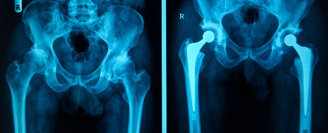
| Hip replacement. Before and after X-rays of a double hip replacement,on a computer screen. The prosthetic hip joints can be clearly seen in the X-ray on the right (after). They consist of a shaft and ball attached to the top of the femur (thigh bone) and a socket (bright semi-circle above the ball) that is inserted into the pelvis. In the X-ray on the right (before),it is difficult to distinguish the joint due to osteoarthritis. Osteoarthritis is a degenerative joint disease that results in the loss of cartilage between the joint and the growth of bone in place of the cartilage. This causes pain,stiffness and loss of mobility | |
| Lizenzart: | Lizenzpflichtig |
| Credit: | Science Photo Library / Reeve, Antonia |
| Bildgröße: | 3985 px × 1616 px |
| Modell-Rechte: | nicht erforderlich |
| Eigentums-Rechte: | nicht erforderlich |
| Restrictions: | - |
Preise für dieses Bild ab 15 €
Universitäten & Organisationen
(Informationsmaterial Digital, Informationsmaterial Print, Lehrmaterial Digital etc.)
ab 15 €
Redaktionell
(Bücher, Bücher: Sach- und Fachliteratur, Digitale Medien (redaktionell) etc.)
ab 30 €
Werbung
(Anzeigen, Aussenwerbung, Digitale Medien, Fernsehwerbung, Karten, Werbemittel, Zeitschriften etc.)
ab 55 €
Handelsprodukte
(bedruckte Textilie, Kalender, Postkarte, Grußkarte, Verpackung etc.)
ab 75 €
Pauschalpreise
Rechtepakete für die unbeschränkte Bildnutzung in Print oder Online
ab 495 €
Keywords
- Arthrose,
- Ball,
- Becken,
- Behandlung,
- Bildschirm,
- Computer,
- Degeneration,
- degenerativ,
- doppelt,
- Duo,
- Einfarbig,
- Ersatz,
- Erwachsene,
- Femur,
- Gelenk,
- Hüfte,
- Hüften,
- Joint,
- Knochen,
- Knorpel,
- Krankheit,
- künstlich,
- Mann,
- Männlich,
- Medizin,
- medizinisch,
- Mensch,
- Monitor,
- OP-Nachsorge,
- Operation,
- Orthopädie,
- Paar,
- pelvin,
- Prothese,
- Radiographie,
- Röntgen,
- Röntgenbild,
- Röntgengerät,
- Schenkel,
- Steckdose,
- vorher,
- Zwei