Knee replacement,X-ray
Bildnummer 11854074
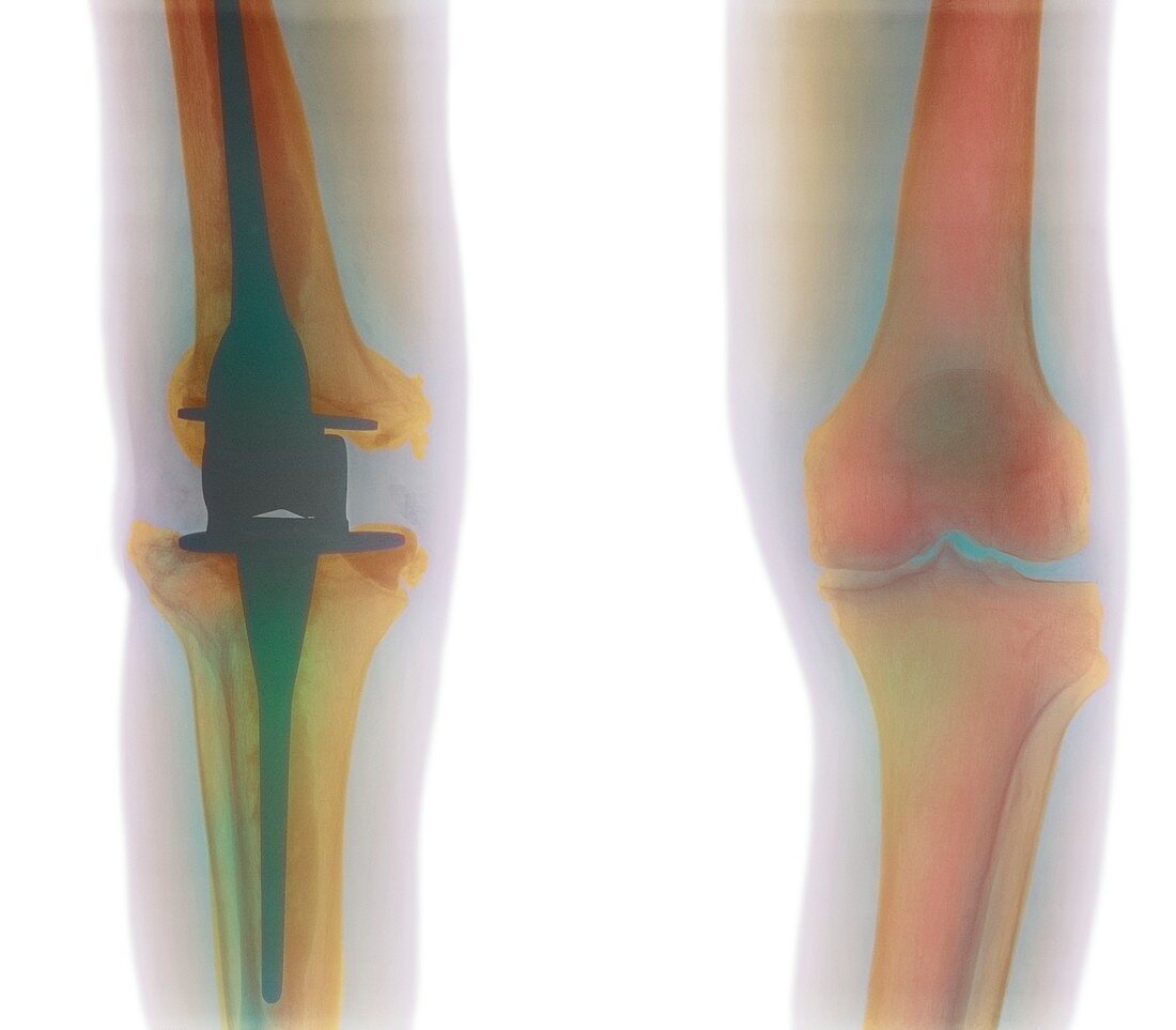
| Knee replacement. Coloured X-ray of the front view of a man's knees,showing a prosthetic knee implant (green,left) and a normal knee (right). The implant is attached by pegs (at upper and lower centre) into the femur and tibia bones (orange) respectively,with surgical cement. The prosthetic joint hinges in an identical way to the normal knee joint. The implant replaced the old joint that had become damaged or lost cartilage,possibly due to an accident or osteoarthritis. Healthy cartilage reduces friction between the bones,and its progressive loss causes joint pain and immobility | |
| Lizenzart: | Lizenzpflichtig |
| Credit: | Science Photo Library |
| Bildgröße: | 3912 px × 3424 px |
| Modell-Rechte: | nicht erforderlich |
| Eigentums-Rechte: | nicht erforderlich |
| Restrictions: | - |
Preise für dieses Bild ab 15 €
Universitäten & Organisationen
(Informationsmaterial Digital, Informationsmaterial Print, Lehrmaterial Digital etc.)
ab 15 €
Redaktionell
(Bücher, Bücher: Sach- und Fachliteratur, Digitale Medien (redaktionell) etc.)
ab 30 €
Werbung
(Anzeigen, Aussenwerbung, Digitale Medien, Fernsehwerbung, Karten, Werbemittel, Zeitschriften etc.)
ab 55 €
Handelsprodukte
(bedruckte Textilie, Kalender, Postkarte, Grußkarte, Verpackung etc.)
ab 75 €
Pauschalpreise
Rechtepakete für die unbeschränkte Bildnutzung in Print oder Online
ab 495 €