Knee replacement,X-ray
Bildnummer 11854062
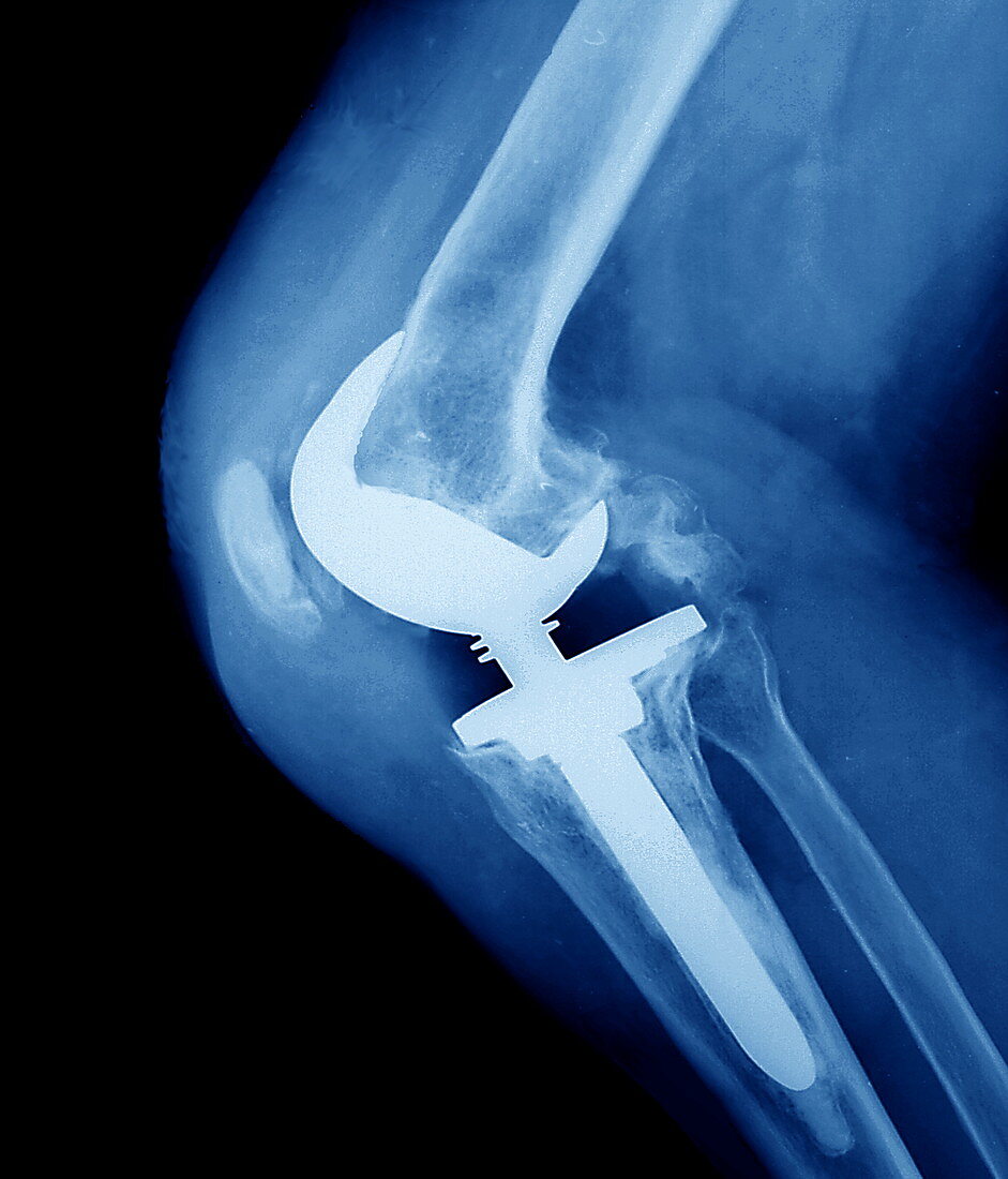
| Knee replacement. Coloured X-ray of the prosthetic knee (white),seen in profile,of a patient with osteoarthritis. The implant attaches to the leg bones (blue/white),and has a flexible joint that can hinge like the old joint. The implant is attached to the top of the tibia (shin bone,lower frame) and to the bottom of the femur (thigh bone,upper frame). The other lower leg bone (fibula) is also seen (right of tibia),as is the patella (kneecap,left of implant). The implant replaced the old joint that had lost its cartilage due to osteoarthritis. Healthy cartilage reduces friction between the bones,and its progressive loss causes joint pain and immobility | |
| Lizenzart: | Lizenzpflichtig |
| Credit: | Science Photo Library / Zephyr |
| Bildgröße: | 3305 px × 3862 px |
| Modell-Rechte: | nicht erforderlich |
| Eigentums-Rechte: | nicht erforderlich |
| Restrictions: | - |
Preise für dieses Bild ab 15 €
Universitäten & Organisationen
(Informationsmaterial Digital, Informationsmaterial Print, Lehrmaterial Digital etc.)
ab 15 €
Redaktionell
(Bücher, Bücher: Sach- und Fachliteratur, Digitale Medien (redaktionell) etc.)
ab 30 €
Werbung
(Anzeigen, Aussenwerbung, Digitale Medien, Fernsehwerbung, Karten, Werbemittel, Zeitschriften etc.)
ab 55 €
Handelsprodukte
(bedruckte Textilie, Kalender, Postkarte, Grußkarte, Verpackung etc.)
ab 75 €
Pauschalpreise
Rechtepakete für die unbeschränkte Bildnutzung in Print oder Online
ab 495 €
Keywords
- Arthritis,
- arthritisch,
- Arthrose,
- Behandlung,
- Bein,
- Biegen,
- Ersatz,
- farbig,
- Femur,
- geduldig,
- Gelenk,
- Gesundheitswesen,
- Joint,
- Knie,
- Kniescheibe,
- Knochen,
- künstliches Implantat,
- Medizin,
- medizinisch,
- monochromes Bild,
- Profil,
- Prothese,
- Radiographie,
- Röntgen,
- Röntgenbild,
- Schenkel,
- Schienbein,
- Schwarz und weiß,
- Seite,
- Wadenbein