Hip replacement
Bildnummer 11854057
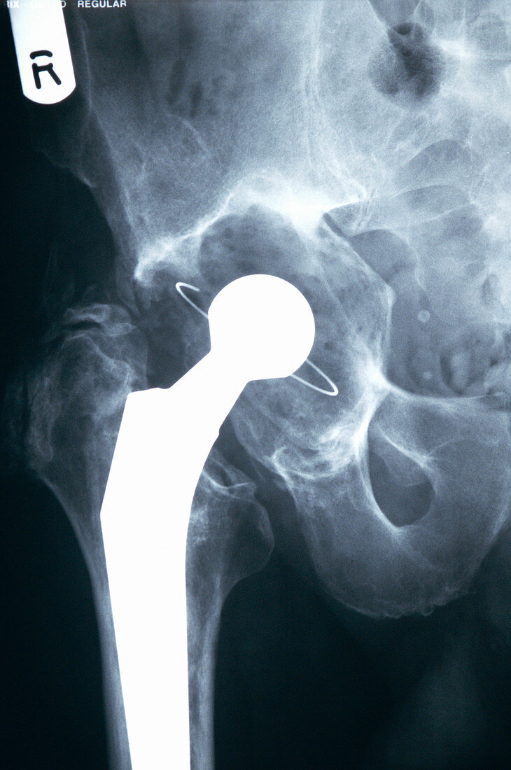
| Hip replacement. X-ray of the leg of a 66-year-old man with a total hip joint replacement (white). The prosthesis consists of a ball and shaft attached to the top of the femur (thigh bone,lower left),and a socket (centre,of which its metal ring is seen),which is fitted into the pelvis. The prosthesis replaced an osteoarthritic hip joint in which the friction-reducing cartilage had been worn away | |
| Lizenzart: | Lizenzpflichtig |
| Credit: | Science Photo Library / Marazzi, Dr. P. |
| Bildgröße: | 3045 px × 4589 px |
| Modell-Rechte: | nicht erforderlich |
| Eigentums-Rechte: | nicht erforderlich |
| Restrictions: | - |
Preise für dieses Bild ab 15 €
Universitäten & Organisationen
(Informationsmaterial Digital, Informationsmaterial Print, Lehrmaterial Digital etc.)
ab 15 €
Redaktionell
(Bücher, Bücher: Sach- und Fachliteratur, Digitale Medien (redaktionell) etc.)
ab 30 €
Werbung
(Anzeigen, Aussenwerbung, Digitale Medien, Fernsehwerbung, Karten, Werbemittel, Zeitschriften etc.)
ab 55 €
Handelsprodukte
(bedruckte Textilie, Kalender, Postkarte, Grußkarte, Verpackung etc.)
ab 75 €
Pauschalpreise
Rechtepakete für die unbeschränkte Bildnutzung in Print oder Online
ab 495 €
Keywords
- Alt,
- älter,
- Arthritis,
- arthritisch,
- Arthrose,
- Becken,
- Bein,
- Einfarbig,
- Femur,
- geduldig,
- Gelenk,
- Gesundheitswesen,
- Greis,
- implantiert,
- Joint,
- künstliches Implantat,
- Mann,
- Männlich,
- Medizin,
- medizinisch,
- OAP,
- Oberschenkelknochen,
- pelvin,
- Prothese,
- rekonstruiert,
- Röntgen,
- Röntgenbild,
- Schwarz und weiß,
- Wiederaufbau