Prosthetic heart valve,3D CT scan
Bildnummer 11853146
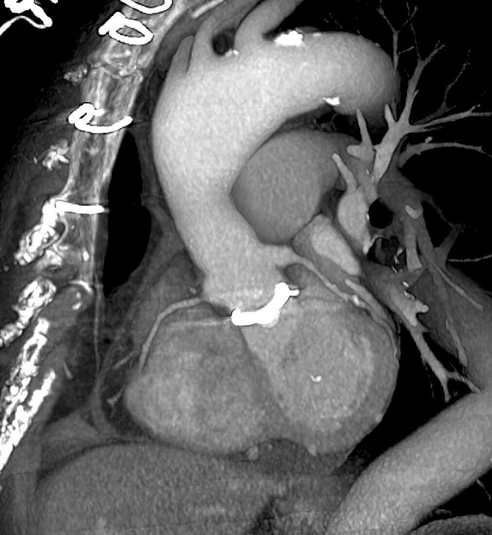
| Prosthetic heart valve. 3D CT (computed tomography) scan of a 70-year-old patient's chest. This is an oblique view of the left side,looking towards the front of the chest (left). The prosthetic heart valve (white) is seen at centre,where the aorta (upper centre) meets the heart (lower centre). The stitches used to close the chest are seen at upper left. The heart valves prevent backflow of blood in the heart. If they start to fail,a condition known as incompetence,then the patient may not get enough oxygenated blood. This leads to shortness of breath,chest pain and possibly a heart attack. The replacement valve here is artificial,but some can be from donors or even from other animals,usually a pig | |
| Lizenzart: | Lizenzpflichtig |
| Credit: | Science Photo Library / Zephyr |
| Bildgröße: | 3648 px × 3969 px |
| Modell-Rechte: | nicht erforderlich |
| Eigentums-Rechte: | nicht erforderlich |
| Restrictions: | - |
Preise für dieses Bild ab 15 €
Universitäten & Organisationen
(Informationsmaterial Digital, Informationsmaterial Print, Lehrmaterial Digital etc.)
ab 15 €
Redaktionell
(Bücher, Bücher: Sach- und Fachliteratur, Digitale Medien (redaktionell) etc.)
ab 30 €
Werbung
(Anzeigen, Aussenwerbung, Digitale Medien, Fernsehwerbung, Karten, Werbemittel, Zeitschriften etc.)
ab 55 €
Handelsprodukte
(bedruckte Textilie, Kalender, Postkarte, Grußkarte, Verpackung etc.)
ab 75 €
Pauschalpreise
Rechtepakete für die unbeschränkte Bildnutzung in Print oder Online
ab 495 €
Keywords
- 3 dimensional,
- 3-d,
- 3-dimensional,
- 3D,
- 70er Jahre,
- Alt,
- älter,
- Aorta,
- Behandlung,
- Computertomographie,
- CT-Scan,
- CT-Scanner,
- Diagnose,
- Dreidimensional,
- Einfarbig,
- Erwachsene,
- Fäden,
- Frau,
- geduldig,
- Gerät,
- Gesundheitswesen,
- Herz,
- Herzarterie,
- Implantat,
- implantiert,
- Kardiologie,
- kardiovaskular,
- Koronararterien,
- künstlich,
- Medizin,
- medizinisch,
- menschlicher Körper,
- Nähte,
- OP-Nachsorge,
- postoperativ,
- Profil,
- Prothese,
- Radiographie,
- Ring,
- Ringe,
- Röntgen,
- Röntgengerät,
- Scanner,
- Schwarz und weiß,
- siebziger Jahre,
- Sutur,
- thorakal,
- Thorax,
- Truhe,
- Ventil,
- Weiblich