Cell viZio imaging system
Bildnummer 11849782
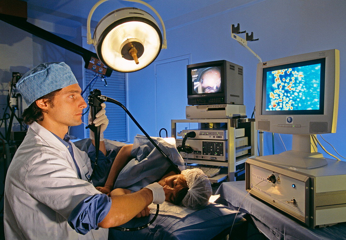
| Cell viZio. Surgeon using the cell viZio imaging system with an endoscope to examine a patient. The cell viZio,which was developed by Mauna Kea Technologies,France,allows microscopic images of tissue to be taken in situ. Both the normal endoscope image (left) and the microscopic image of the tissue (right) can be seen. The cell viZio allows diagnosis of diseases such as cancer by 'optical biopsy',a minimally invasive technique. The system consists of a long tube containing thousands of optical fibres. At the tip of the tube is a very thin probe (350 microns diameter),which enters the tissue. A laser shines on the tissue and relays the microscopic image to a computer,which displays the image on a monitor | |
| Lizenzart: | Lizenzpflichtig |
| Credit: | Science Photo Library / Psaila, Philippe |
| Bildgröße: | 5030 px × 3487 px |
| Modell-Rechte: | Derzeit liegt noch kein Release vor. Bitte kontaktieren Sie uns vor Verwendung. |
| Eigentums-Rechte: | nicht erforderlich |
| Restrictions: | - |
Preise für dieses Bild ab 15 €
Universitäten & Organisationen
(Informationsmaterial Digital, Informationsmaterial Print, Lehrmaterial Digital etc.)
ab 15 €
Redaktionell
(Bücher, Bücher: Sach- und Fachliteratur, Digitale Medien (redaktionell) etc.)
ab 30 €
Werbung
(Anzeigen, Aussenwerbung, Digitale Medien, Fernsehwerbung, Karten, Werbemittel, Zeitschriften etc.)
ab 55 €
Handelsprodukte
(bedruckte Textilie, Kalender, Postkarte, Grußkarte, Verpackung etc.)
ab 75 €
Pauschalpreise
Rechtepakete für die unbeschränkte Bildnutzung in Print oder Online
ab 495 €
Keywords
- 20er Jahre,
- 30er Jahre,
- Arzt,
- Begutachten,
- Bildschirm,
- Bildschirme,
- Cell Vizio,
- Chirurg,
- chirurgisch,
- Computer,
- Diagnose,
- dreißiger Jahre,
- Endoskop,
- Erwachsene,
- fcfm,
- Fiberoptik,
- flexibel,
- Französisch,
- geduldig,
- Gerät,
- Histologie,
- histologisch,
- Instrument,
- Kamera,
- kaukasisch,
- Krankenhaus,
- Laser,
- Mann,
- Männlich,
- Maschine,
- Mauna Kea Technologien,
- Medizin,
- medizinisch,
- Mensch,
- Mikroskopie,
- minimal-invasive,
- Monitor,
- Monitore,
- Operation,
- Operationssaal,
- optische Biopsie,
- Sonde,
- System,
- Technologie,
- technologisch,
- Überprüfung,
- Untersuchung,
- zwanziger Jahre