Patient undergoing a PET scan of the brain
Bildnummer 11848862
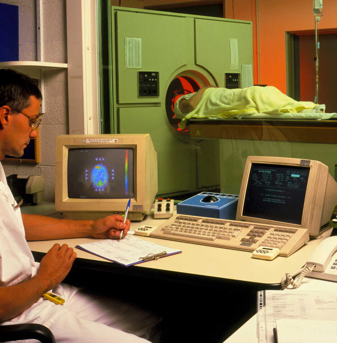
| MODEL RELEASED. PET scan. Technician studying a positron emission tomography (PET) scan of a patient's brain. This is a technique used to detect areas of abnormal activity in the body. The result of one scan is seen on the monitor at centre left. The brain shows an area of high activity in the left frontal lobe (red). Prior to the scan,radioactive glucose is injected into the bloodstream. The PET scanner detects this,and builds up a picture of glucose concentration,and thus metabolic activity,in the scanned area. This is particularly useful in detecting brain tumours,which show a marked difference in activity to healthy brain tissue | |
| Lizenzart: | Lizenzpflichtig |
| Credit: | Science Photo Library / CC Studio |
| Bildgröße: | 3524 px × 3578 px |
| Modell-Rechte: | vorhanden |
| Eigentums-Rechte: | nicht erforderlich |
| Restrictions: | - |
Preise für dieses Bild ab 15 €
Universitäten & Organisationen
(Informationsmaterial Digital, Informationsmaterial Print, Lehrmaterial Digital etc.)
ab 15 €
Redaktionell
(Bücher, Bücher: Sach- und Fachliteratur, Digitale Medien (redaktionell) etc.)
ab 30 €
Werbung
(Anzeigen, Aussenwerbung, Digitale Medien, Fernsehwerbung, Karten, Werbemittel, Zeitschriften etc.)
ab 55 €
Handelsprodukte
(bedruckte Textilie, Kalender, Postkarte, Grußkarte, Verpackung etc.)
ab 75 €
Pauschalpreise
Rechtepakete für die unbeschränkte Bildnutzung in Print oder Online
ab 495 €