3-D CT scan of infant with deformed skull
Bildnummer 11845852
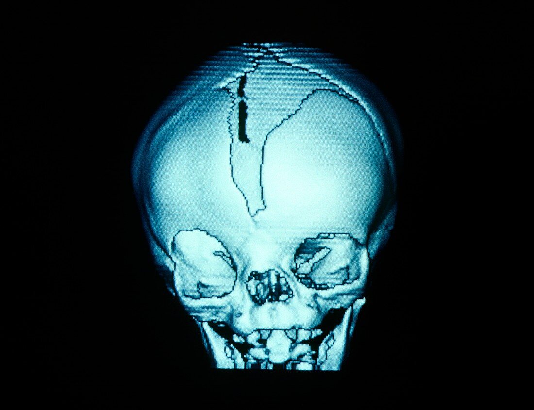
| Monochrome 3-D image showing a frontal view of the skull of an infant suffering from a skull growth abnormality. The image is one of a number of computer reconstructions from a series of conventional computed tomography (CT) scans that were used in planning & simulating the necessary corrective surgery. In a normal infant,vertical grooves in the skull (the cranial sutures) remain open throughout the 1st year of life,acting as expansion joints for the growing brain. In this case,a fused suture has caused uneven growth. This image reveals facial asymmetry. The right eye socket (orbit) on left of image is vertically enlarged,compared to the left orbit | |
| Lizenzart: | Lizenzpflichtig |
| Credit: | Science Photo Library / Tsiaras, Alexander |
| Bildgröße: | 4827 px × 3709 px |
| Modell-Rechte: | nicht erforderlich |
| Eigentums-Rechte: | nicht erforderlich |
| Restrictions: |
|
Preise für dieses Bild ab 15 €
Universitäten & Organisationen
(Informationsmaterial Digital, Informationsmaterial Print, Lehrmaterial Digital etc.)
ab 15 €
Redaktionell
(Bücher, Bücher: Sach- und Fachliteratur, Digitale Medien (redaktionell) etc.)
ab 30 €
Werbung
(Anzeigen, Aussenwerbung, Digitale Medien, Fernsehwerbung, Karten, Werbemittel, Zeitschriften etc.)
ab 55 €
Handelsprodukte
(bedruckte Textilie, Kalender, Postkarte, Grußkarte, Verpackung etc.)
ab 75 €
Pauschalpreise
Rechtepakete für die unbeschränkte Bildnutzung in Print oder Online
ab 495 €