3-D CT scans of infant skull post-op for deformity
Bildnummer 11845849
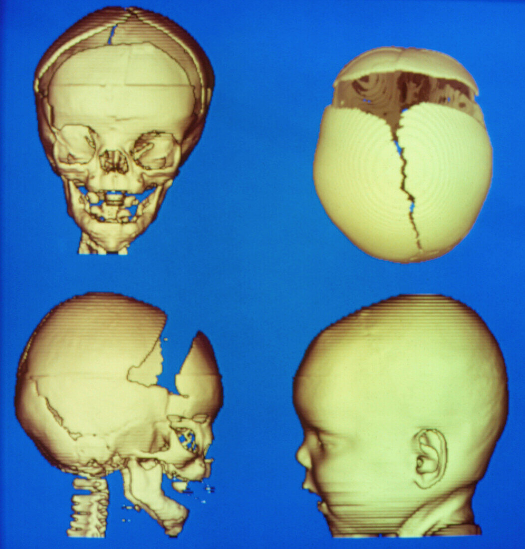
| 3-D reconstructions from conventional computed tomography (CT) scans showing the post-operative appearance of an infant's skull following surgery to correct a growth abnormality. The blue defect in the bottom left scan represents a section of bone from the right forehead removed by the surgeon; the surrounding bones will close naturally in time. In a normal infant,grooves running down the skull act as expansion joints during the first year to accommodate a growing brain. In this child,a fused groove (cranial suture) was causing uneven growth & progressive deformity. Various 3-D reconstructions were used in planning & simulating the operation | |
| Lizenzart: | Lizenzpflichtig |
| Credit: | Science Photo Library / Tsiaras, Alexander |
| Bildgröße: | 3390 px × 3543 px |
| Modell-Rechte: | nicht erforderlich |
| Eigentums-Rechte: | nicht erforderlich |
| Restrictions: |
|
Preise für dieses Bild ab 15 €
Universitäten & Organisationen
(Informationsmaterial Digital, Informationsmaterial Print, Lehrmaterial Digital etc.)
ab 15 €
Redaktionell
(Bücher, Bücher: Sach- und Fachliteratur, Digitale Medien (redaktionell) etc.)
ab 30 €
Werbung
(Anzeigen, Aussenwerbung, Digitale Medien, Fernsehwerbung, Karten, Werbemittel, Zeitschriften etc.)
ab 55 €
Handelsprodukte
(bedruckte Textilie, Kalender, Postkarte, Grußkarte, Verpackung etc.)
ab 75 €
Pauschalpreise
Rechtepakete für die unbeschränkte Bildnutzung in Print oder Online
ab 495 €