Spondylolisthesis back deformity,MRI
Bildnummer 11844844
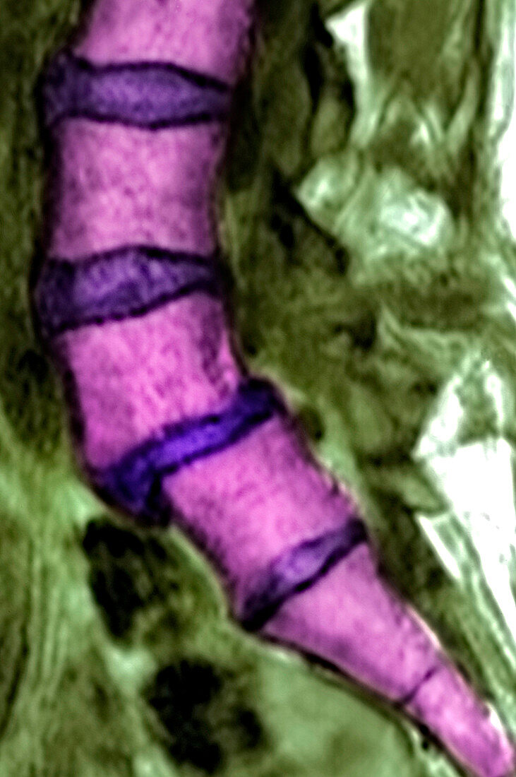
| Spondylolisthesis,image 1 of 2. Coloured magnetic resonance imaging (MRI) scan of a side view of the lower back of a 46 year old woman showing spondylolisthesis. This is the backward displacement of a vertebra (spinal bone,light purple) relative to the one above it. Here it is between the lumbar vertebrae L4 (centre left) and L5 (lower centre). The condition is often due to a hereditary bone disorder,but can also be caused by trauma. It typically causes back pain,and the displaced vertebra puts pressure on the nearby spinal cord,causing sciatica (leg pain),loss of bladder and/or bowel control and eventually paralysis of the lower limbs. For the same patient after five years,see image M330/1432 | |
| Lizenzart: | Lizenzpflichtig |
| Credit: | Science Photo Library / Marazzi, Dr. P. |
| Bildgröße: | 3408 px × 5130 px |
| Modell-Rechte: | nicht erforderlich |
| Eigentums-Rechte: | nicht erforderlich |
| Restrictions: | - |
Preise für dieses Bild ab 15 €
Universitäten & Organisationen
(Informationsmaterial Digital, Informationsmaterial Print, Lehrmaterial Digital etc.)
ab 15 €
Redaktionell
(Bücher, Bücher: Sach- und Fachliteratur, Digitale Medien (redaktionell) etc.)
ab 30 €
Werbung
(Anzeigen, Aussenwerbung, Digitale Medien, Fernsehwerbung, Karten, Werbemittel, Zeitschriften etc.)
ab 55 €
Handelsprodukte
(bedruckte Textilie, Kalender, Postkarte, Grußkarte, Verpackung etc.)
ab 75 €
Pauschalpreise
Rechtepakete für die unbeschränkte Bildnutzung in Print oder Online
ab 495 €
Keywords
- 40er Jahre,
- Bandscheiben,
- Deformität,
- Diagnose,
- Erwachsene,
- farbig,
- Frau,
- früh,
- geduldig,
- gefärbt,
- Gesundheitswesen,
- Knochen,
- Kondition,
- L4,
- L5,
- Lendenwirbelsäule,
- Magnetresonanztomografie,
- Medizin,
- medizinisch,
- menschlicher Körper,
- MRT-Untersuchung,
- Profil,
- Rückenschmerzen,
- Scanner,
- schmerzhaft,
- Seitenansicht,
- spondylolisthesis,
- Störung,
- unterer Rücken,
- vertebral,
- Vierziger Jahre,
- Weiblich,
- Wirbel,
- Wirbelsäule