Pinned ankle fractures,X-ray
Bildnummer 11844811
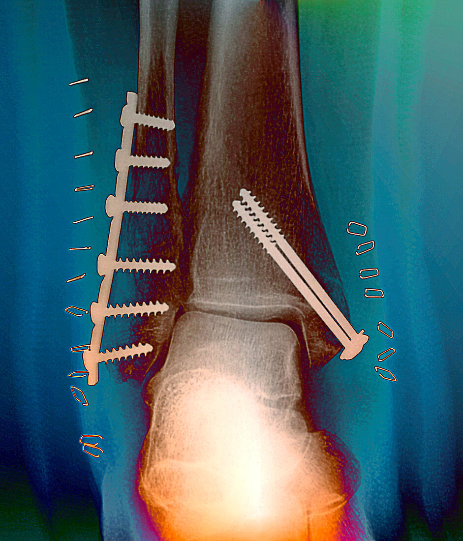
| Pinned ankle fracture. Coloured frontal X-ray of the ankle bones of a patient with pinned ankle fractures. The malleolus (bony projection at the end of a bone) of both lower leg bones have been fractured and then pinned back together. The fibula (left) is the thinner of the two lower leg bones,and has had a plate as well as screws used to hold it together. The pins and plates hold the bone fragments in position while they heal. The tibia (right) is the thicker of the two lower leg bones. The bones at bottom are part of the ankle bones. Staples are seen in the surrounding tissue,showing where the skin was stapled back together following the bone-pinning operation. This is the patient's right ankle | |
| Lizenzart: | Lizenzpflichtig |
| Credit: | Science Photo Library / Zephyr |
| Bildgröße: | 3402 px × 3976 px |
| Modell-Rechte: | nicht erforderlich |
| Eigentums-Rechte: | nicht erforderlich |
| Restrictions: | - |
Preise für dieses Bild ab 15 €
Universitäten & Organisationen
(Informationsmaterial Digital, Informationsmaterial Print, Lehrmaterial Digital etc.)
ab 15 €
Redaktionell
(Bücher, Bücher: Sach- und Fachliteratur, Digitale Medien (redaktionell) etc.)
ab 30 €
Werbung
(Anzeigen, Aussenwerbung, Digitale Medien, Fernsehwerbung, Karten, Werbemittel, Zeitschriften etc.)
ab 55 €
Handelsprodukte
(bedruckte Textilie, Kalender, Postkarte, Grußkarte, Verpackung etc.)
ab 75 €
Pauschalpreise
Rechtepakete für die unbeschränkte Bildnutzung in Print oder Online
ab 495 €
Keywords
- anterior,
- Ausrüstung,
- Behandlung,
- Bein,
- farbig,
- festgesteckt,
- Fraktur,
- frakturiert,
- Frontal,
- geduldig,
- gefärbt,
- Gelenk,
- Gesundheitswesen,
- Implantat,
- Joint,
- kaputt,
- Knöchel,
- Knochen,
- Kondition,
- Medizin,
- medizinisch,
- menschlicher Körper,
- Nadel,
- postoperativ,
- Radiographie,
- Recht,
- repariert,
- Röntgen,
- Röntgengerät,
- Schraube,
- Skelett-,
- Störung,
- Technologie,
- technologisch,
- Teller,
- Unterbrechung,
- verletzt,
- Verletzung