Slipped disc,MRI
Bildnummer 11844485
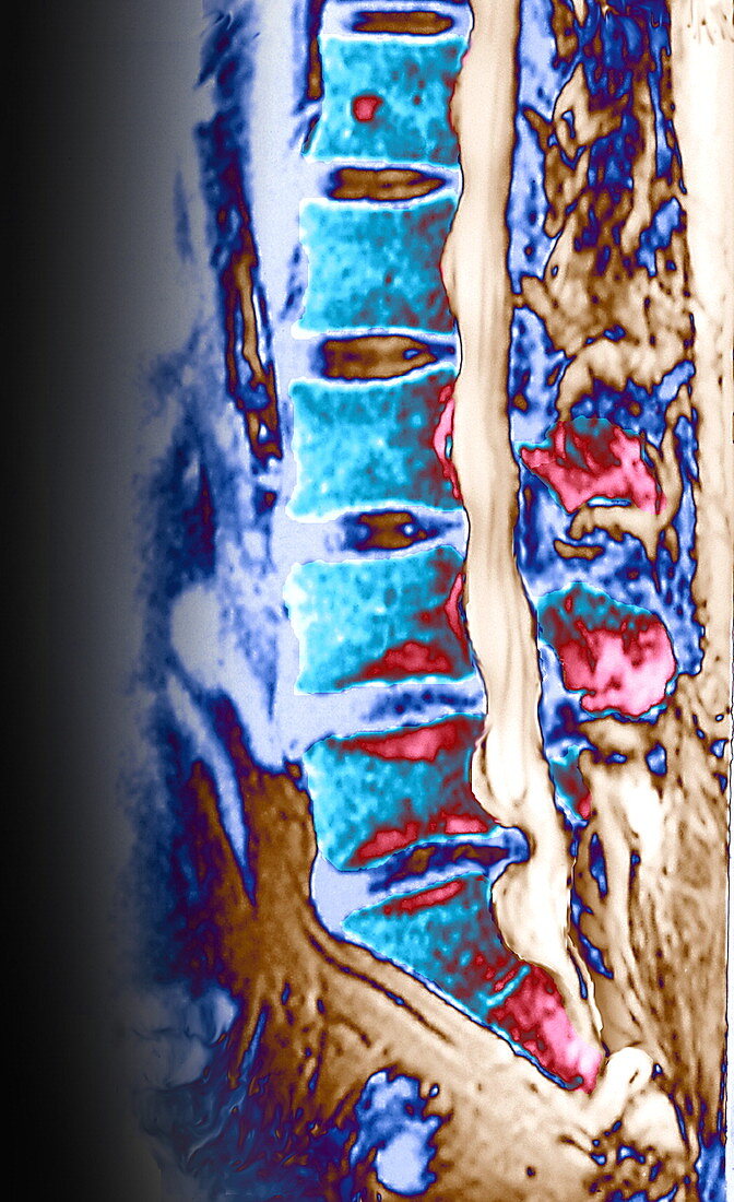
| Slipped disc. Coloured magnetic resonance imaging (MRI) scan of a sagittal (side) section through the lower spine of a patient suffering from a slipped or herniated disc. The central part (dark blue,lower right) of one of the discs that separate the bones (vertebrae,light blue) of the spine has been forced through a weakened area of the disc,and is protruding into the spinal cord (beige strip,top to lower right). A slipped disc often occurs after a back injury or strain,causing back pain. It may compress the nerves of the spinal cord,resulting in tingling or numbness in the feet and legs. Treatment includes rest,anti-inflammatory drugs and physical therapy | |
| Lizenzart: | Lizenzpflichtig |
| Credit: | Science Photo Library / GJLP |
| Bildgröße: | 1039 px × 1701 px |
| Modell-Rechte: | nicht erforderlich |
| Eigentums-Rechte: | nicht erforderlich |
| Restrictions: | - |
Preise für dieses Bild ab 15 €
Universitäten & Organisationen
(Informationsmaterial Digital, Informationsmaterial Print, Lehrmaterial Digital etc.)
ab 15 €
Redaktionell
(Bücher, Bücher: Sach- und Fachliteratur, Digitale Medien (redaktionell) etc.)
ab 30 €
Werbung
(Anzeigen, Aussenwerbung, Digitale Medien, Fernsehwerbung, Karten, Werbemittel, Zeitschriften etc.)
ab 55 €
Handelsprodukte
(bedruckte Textilie, Kalender, Postkarte, Grußkarte, Verpackung etc.)
ab 75 €
Pauschalpreise
Rechtepakete für die unbeschränkte Bildnutzung in Print oder Online
ab 495 €