Spondylitis of spine,MRI
Bildnummer 11843084
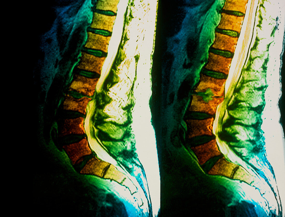
| Spondylitis. Coloured magnetic resonance imaging (MRI) scans of sagittal (side) sections through the lower spine of a 63-year-old patient suffering from spondylitis. The front of the body is at left in both. Intervertebral discs (green) separate the lumbar vertebrae (bones,orange). The space between vertebrae L2 and L3 (centre left and right) has narrowed,due to inflammation of the joint (spondylitis). Part of the joint is pressing on the spinal cord (white,to the right of the vertebrae). Spondylitis causes pain and restricts movement. Treatment includes physical therapy and anti-inflammatory drugs. In severe cases,surgery can relieve pressure on the spinal canal | |
| Lizenzart: | Lizenzpflichtig |
| Credit: | Science Photo Library / Zephyr |
| Bildgröße: | 4771 px × 3641 px |
| Modell-Rechte: | nicht erforderlich |
| Eigentums-Rechte: | nicht erforderlich |
| Restrictions: | - |
Preise für dieses Bild ab 15 €
Universitäten & Organisationen
(Informationsmaterial Digital, Informationsmaterial Print, Lehrmaterial Digital etc.)
ab 15 €
Redaktionell
(Bücher, Bücher: Sach- und Fachliteratur, Digitale Medien (redaktionell) etc.)
ab 30 €
Werbung
(Anzeigen, Aussenwerbung, Digitale Medien, Fernsehwerbung, Karten, Werbemittel, Zeitschriften etc.)
ab 55 €
Handelsprodukte
(bedruckte Textilie, Kalender, Postkarte, Grußkarte, Verpackung etc.)
ab 75 €
Pauschalpreise
Rechtepakete für die unbeschränkte Bildnutzung in Print oder Online
ab 495 €
Keywords
- Abschnitte,
- Alt,
- älter,
- Bandscheiben,
- Bild,
- Bilder,
- Degeneration,
- entzündet,
- Entzündung,
- farbig,
- Gelenk,
- Gelenke,
- Gesundheitswesen,
- Greis,
- Joint,
- Kanal,
- Knochen,
- Kondition,
- Krankheit,
- L2,
- L3,
- Lendenwirbelsäule,
- Magnetresonanztomografie,
- Medizin,
- medizinisch,
- MRI,
- niedriger,
- OAP,
- Pensionäre,
- Rentner,
- Rücken,
- Rückgrat,
- Säule,
- Scan,
- Scannen,
- Seite,
- Sektion,
- sektioniert,
- Spondylitis,
- Störung,
- vertebral,
- Wirbel,
- Wirbelsäule,
- Wirbelsäulen-