Coloured MRI brain scan of Sturge-Weber syndrome
Bildnummer 11843057
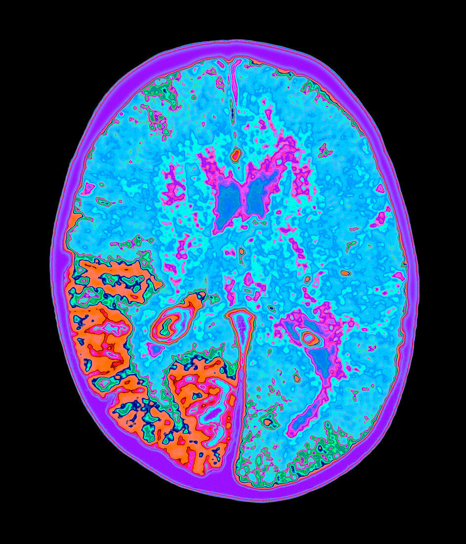
| Sturge-Weber syndrome. Coloured magnetic resonance imaging (MRI) scan of a brain (light blue/orange) with Sturge-Weber syndrome. The brain is seen in horizontal (axial) section,with the front of the brain at top. The brain's fluid-filled ventricles are dark blue. Part of the brain has become calci- fied (orange,lower left). Other symptoms include angioma tumours in the choroid of the eye and the meninges membranes that cover the brain,as well as port-wine nevi on the face and glaucoma of the eye. People with this congenital disorder are mentally handicapped and may suffer from epileptic seizures. MRI uses radio waves and a powerful magnet to produce slice images of the body | |
| Lizenzart: | Lizenzpflichtig |
| Credit: | Science Photo Library / Kulyk, Mehau |
| Bildgröße: | 3000 px × 3500 px |
| Modell-Rechte: | nicht erforderlich |
| Eigentums-Rechte: | nicht erforderlich |
| Restrictions: | - |
Preise für dieses Bild ab 15 €
Universitäten & Organisationen
(Informationsmaterial Digital, Informationsmaterial Print, Lehrmaterial Digital etc.)
ab 15 €
Redaktionell
(Bücher, Bücher: Sach- und Fachliteratur, Digitale Medien (redaktionell) etc.)
ab 30 €
Werbung
(Anzeigen, Aussenwerbung, Digitale Medien, Fernsehwerbung, Karten, Werbemittel, Zeitschriften etc.)
ab 55 €
Handelsprodukte
(bedruckte Textilie, Kalender, Postkarte, Grußkarte, Verpackung etc.)
ab 75 €
Pauschalpreise
Rechtepakete für die unbeschränkte Bildnutzung in Print oder Online
ab 495 €