Brain Scan
Bildnummer 11842239
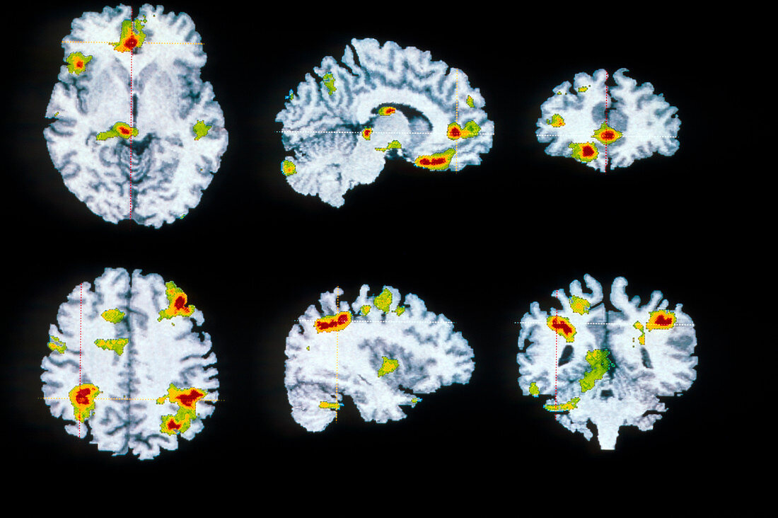
| Obsessive-compulsive disorder. Coloured Positron Emission Tomography (PET) scans of a human brain,showing active areas in obsessive compulsive disorder. The brain is sectioned axially (left),sagittally (centre),coronally (at right). In this patient,positive correlations (activity increases as symptoms get stronger) are in the top row,seen coloured in the left orbital region,prefrontal,left frontal gyri & thalamus. Negative correlation (activity decreasing as symptoms strengthen) are in the bottom row in the right frontal gyrus and parietal regions. Active areas coloured red/yellow show blood flow detected by a radioactive tracer | |
| Lizenzart: | Lizenzpflichtig |
| Credit: | Science Photo Library / Wellcome Dept. of Cognitive Neurology |
| Bildgröße: | 3777 px × 2515 px |
| Modell-Rechte: | nicht erforderlich |
| Eigentums-Rechte: | nicht erforderlich |
| Restrictions: | - |
Preise für dieses Bild ab 15 €
Universitäten & Organisationen
(Informationsmaterial Digital, Informationsmaterial Print, Lehrmaterial Digital etc.)
ab 15 €
Redaktionell
(Bücher, Bücher: Sach- und Fachliteratur, Digitale Medien (redaktionell) etc.)
ab 30 €
Werbung
(Anzeigen, Aussenwerbung, Digitale Medien, Fernsehwerbung, Karten, Werbemittel, Zeitschriften etc.)
ab 55 €
Handelsprodukte
(bedruckte Textilie, Kalender, Postkarte, Grußkarte, Verpackung etc.)
ab 75 €
Pauschalpreise
Rechtepakete für die unbeschränkte Bildnutzung in Print oder Online
ab 495 €