Pneumothorax,3D CT scan
Bildnummer 11842097
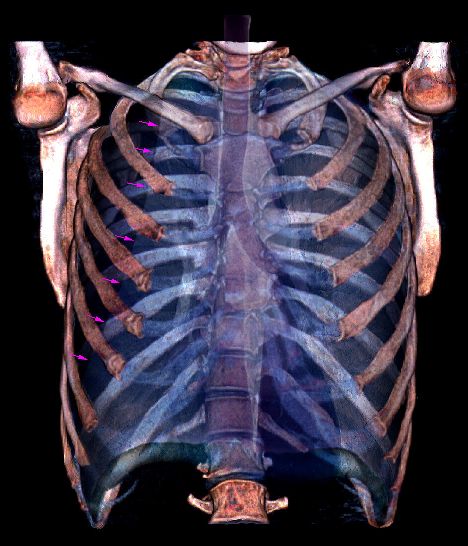
| Pneumothorax. Coloured frontal 3D computed tomography (CT) scan of the lungs of a patient with a pneumothorax,or collapsed lung. The arrows down left show where the right lung has collapsed to leave a dark,air-filled space. A pneumothorax occurs due to trauma or lung rupture. It causes shortness of breath and pain on breathing. A pneumothorax can be treated by inserting a tube into the air-filled space and draining the air over a period of days. This allows the collapsed lung to re-inflate. The structures seen inside the rib cage are the trachea (windpipe),running from the throat (top centre) down to centre,where it splits into the two bronchi,one for each lung (blue spaces at left and right) | |
| Lizenzart: | Lizenzpflichtig |
| Credit: | Science Photo Library / Zephyr |
| Bildgröße: | 3402 px × 3969 px |
| Modell-Rechte: | nicht erforderlich |
| Eigentums-Rechte: | nicht erforderlich |
| Restrictions: | - |
Preise für dieses Bild ab 15 €
Universitäten & Organisationen
(Informationsmaterial Digital, Informationsmaterial Print, Lehrmaterial Digital etc.)
ab 15 €
Redaktionell
(Bücher, Bücher: Sach- und Fachliteratur, Digitale Medien (redaktionell) etc.)
ab 30 €
Werbung
(Anzeigen, Aussenwerbung, Digitale Medien, Fernsehwerbung, Karten, Werbemittel, Zeitschriften etc.)
ab 55 €
Handelsprodukte
(bedruckte Textilie, Kalender, Postkarte, Grußkarte, Verpackung etc.)
ab 75 €
Pauschalpreise
Rechtepakete für die unbeschränkte Bildnutzung in Print oder Online
ab 495 €
Keywords
- 3-d,
- 3D,
- anterior,
- Atmungssystem,
- Computertomographie,
- CT-Scan,
- CT-Scanner,
- Diagnose,
- Dreidimensional,
- Erwachsene,
- farbig,
- Frontal,
- geduldig,
- gefärbt,
- Gesundheitswesen,
- kollabierte Lunge,
- Kondition,
- Lunge,
- Lungen,
- Medizin,
- medizinisch,
- menschlicher Körper,
- Organ,
- pulmonal,
- Scanner,
- Störung,
- Thorax,
- Truhe