Brain response in obesity,PET scans
Bildnummer 11841558
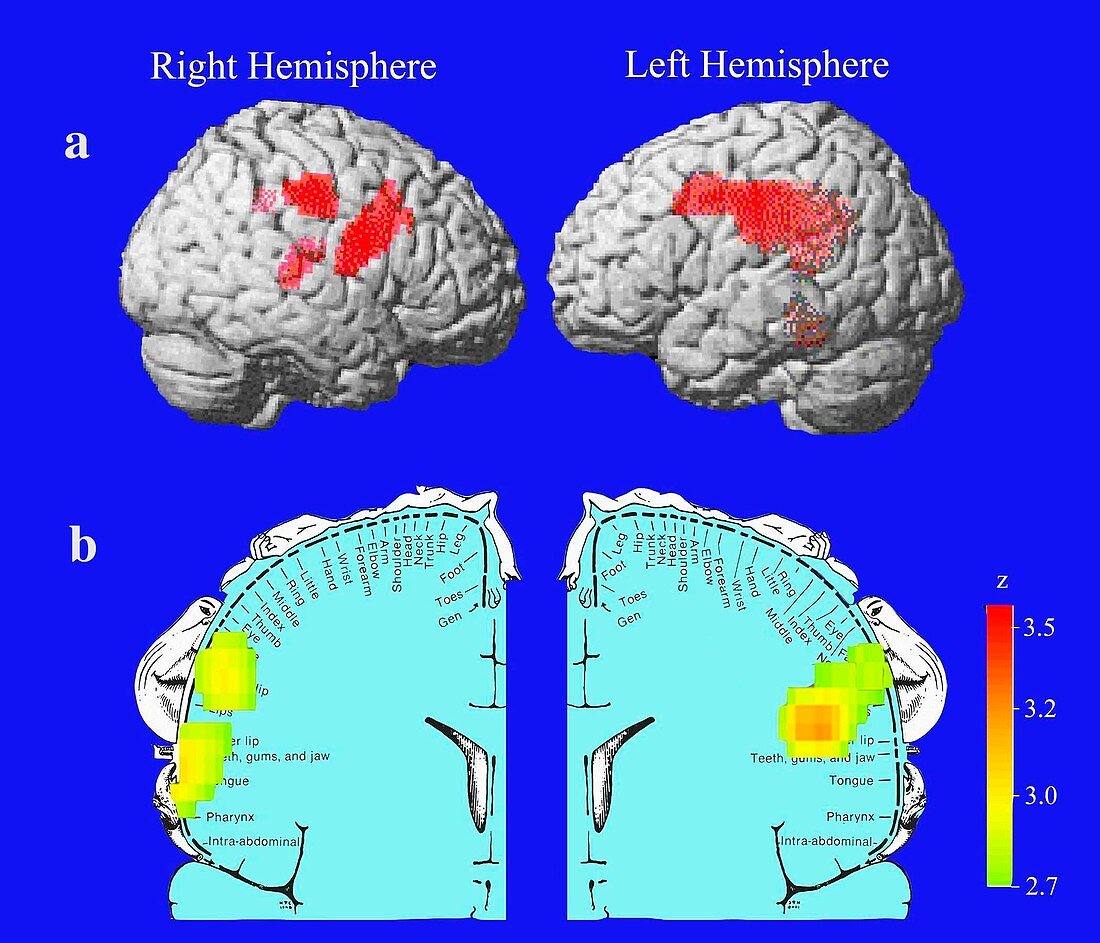
| Brain activity in obesity illustrated by positron emission tomography (PET) scan data overlaid on models of the brain. Anatomical scans (top) of the right and left brain hemispheres (in profile and facing each other) show areas (red) of the brain with increased sensory activity in obese people. A view from the front (bottom) shows a diagram of a coronal (frontal) section through a brain. This shows the somatosensory cortex (the brain's outer layer) where sensory inputs are received from the body. Increased sensitivity (yellow) of the mouth,lips and tongue may help explain obesity. Results presented in 2002 by Gene-Jack Wang | |
| Lizenzart: | Lizenzpflichtig |
| Credit: | Science Photo Library / Brookhaven National Laboratory |
| Bildgröße: | 2100 px × 1800 px |
| Modell-Rechte: | nicht erforderlich |
| Eigentums-Rechte: | nicht erforderlich |
| Restrictions: |
|
Preise für dieses Bild ab 15 €
Universitäten & Organisationen
(Informationsmaterial Digital, Informationsmaterial Print, Lehrmaterial Digital etc.)
ab 15 €
Redaktionell
(Bücher, Bücher: Sach- und Fachliteratur, Digitale Medien (redaktionell) etc.)
ab 30 €
Werbung
(Anzeigen, Aussenwerbung, Digitale Medien, Fernsehwerbung, Karten, Werbemittel, Zeitschriften etc.)
ab 55 €
Handelsprodukte
(bedruckte Textilie, Kalender, Postkarte, Grußkarte, Verpackung etc.)
ab 75 €
Pauschalpreise
Rechtepakete für die unbeschränkte Bildnutzung in Print oder Online
ab 495 €
Keywords
- adipös,
- Adipositas,
- Aktivität,
- cerebral,
- Diagramm,
- Experiment,
- farbcodiert,
- farbig,
- Fett,
- Forschung,
- Gehirn,
- Gesundheitswesen,
- Hemisphären,
- Illustration,
- Karte,
- Kunstwerk,
- Lebensmittel,
- Medizin,
- medizinisch,
- metabolisch,
- Neuroimaging,
- nuklear,
- Physiologie,
- physiologisch,
- Stoffwechsel,
- Sucht,
- Übergewicht,
- Vergleich,
- vergleichen,
- Wang