Inflamed lymph node,Doppler ultrasound
Bildnummer 11840860
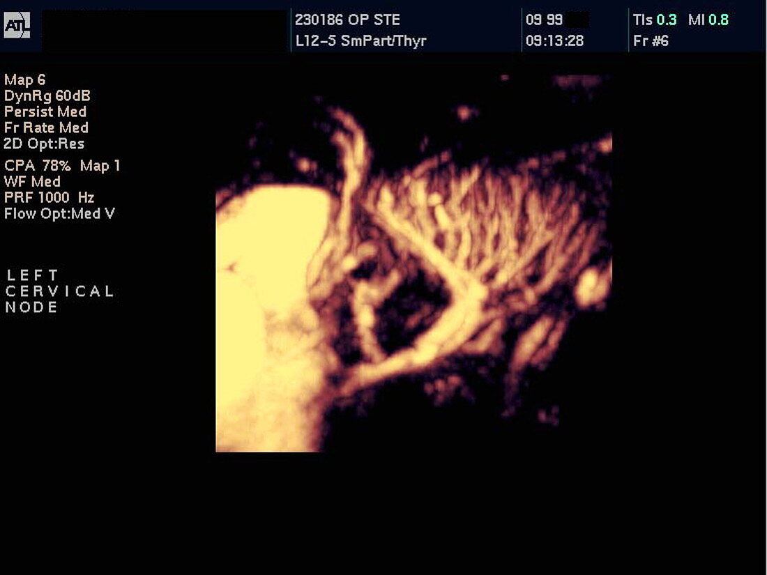
| Inflamed lymph node. Three-dimensional power Doppler ultrasound scan of blood vessels (orange) in an inflamed lymph node in the neck of a 13- year-old boy. The node contains a network of thin blood vessels (centre right). These drain into the thicker jugular vein at left. Inflammation of a lymph node (lymphadenitis) is usually a response to an infection. The increased lymph node activity produces more infection-fighting lymphocyte cells. Doppler ultrasound scanning detects moving fluids,such as blood,by the frequency change they cause when reflecting high-frequency sound waves. Power Doppler scans use colour to show the moving fluid,but do not show the direction of the flow | |
| Lizenzart: | Lizenzpflichtig |
| Credit: | Science Photo Library / Fraser, Simon |
| Bildgröße: | 768 px × 576 px |
| Modell-Rechte: | nicht erforderlich |
| Eigentums-Rechte: | nicht erforderlich |
| Restrictions: | - |
Preise für dieses Bild ab 15 €
Universitäten & Organisationen
(Informationsmaterial Digital, Informationsmaterial Print, Lehrmaterial Digital etc.)
ab 15 €
Redaktionell
(Bücher, Bücher: Sach- und Fachliteratur, Digitale Medien (redaktionell) etc.)
ab 30 €
Werbung
(Anzeigen, Aussenwerbung, Digitale Medien, Fernsehwerbung, Karten, Werbemittel, Zeitschriften etc.)
ab 55 €
Handelsprodukte
(bedruckte Textilie, Kalender, Postkarte, Grußkarte, Verpackung etc.)
ab 75 €
Pauschalpreise
Rechtepakete für die unbeschränkte Bildnutzung in Print oder Online
ab 495 €
Keywords
- 3-d,
- 3-dimensional,
- Blut,
- Diagnose,
- Drei,
- Dreidimensional,
- Drüse,
- entzündet,
- Entzündung,
- Farbe,
- Fließen,
- geduldig,
- Gefäß,
- Gefäße,
- geschwollen,
- Gesundheitswesen,
- Hals,
- Halsader,
- immun,
- Infektion,
- infiziert,
- Junge,
- Kind,
- Kondition,
- Krankheit,
- Kreislauf,
- Leistung,
- Lymphknoten,
- Lymphsystem,
- Männlich,
- Medizin,
- medizinisch,
- Pädiatrie,
- pädiatrisch,
- Scan,
- Scannen,
- Schwellung,
- Sonographie,
- Störung,
- System,
- Teenager,
- Teenageralter,
- Ultraschall,
- vaskulär,
- Vene