Intravenous pyelogram showing duplex kidney
Bildnummer 11840646
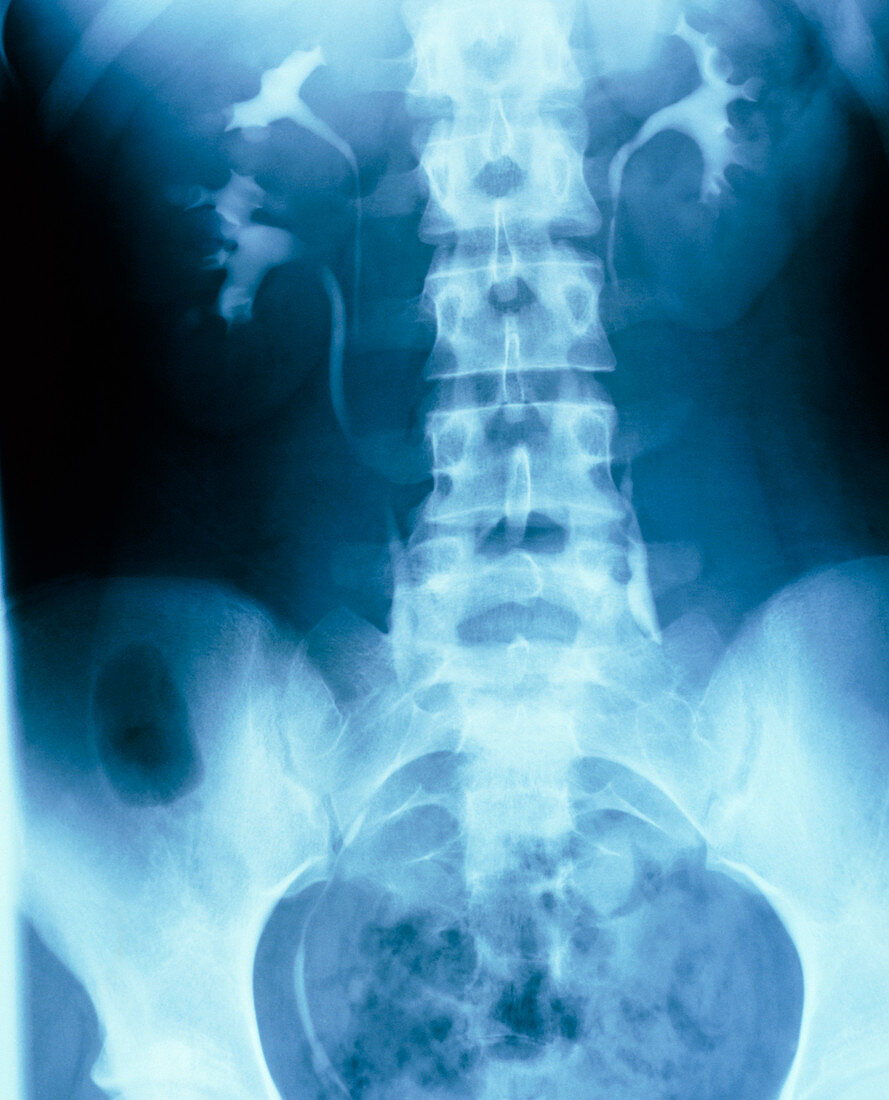
| Duplex kidney: intravenous pyelogram (IVP),an X- ray image of the human abdominal area which displays the drainage of urine from both kidneys down the ureters towards the bladder. Here,the right kidney (left on image) is partially duplicated; two calyces give rise to separate ureters. In IVP,an iodine-based contrast medium,excreted by the kidneys in urine,is injected intravenously prior to the X-ray being taken. The branched calyces - the collecting ducts of the kidneys - & the ureters draining down to the bladder (at bottom,in basin of pelvis) are traced as opaque highlights on the X-ray | |
| Lizenzart: | Lizenzpflichtig |
| Credit: | Science Photo Library |
| Bildgröße: | 3784 px × 4680 px |
| Modell-Rechte: | nicht erforderlich |
| Eigentums-Rechte: | nicht erforderlich |
| Restrictions: | - |
Preise für dieses Bild ab 15 €
Universitäten & Organisationen
(Informationsmaterial Digital, Informationsmaterial Print, Lehrmaterial Digital etc.)
ab 15 €
Redaktionell
(Bücher, Bücher: Sach- und Fachliteratur, Digitale Medien (redaktionell) etc.)
ab 30 €
Werbung
(Anzeigen, Aussenwerbung, Digitale Medien, Fernsehwerbung, Karten, Werbemittel, Zeitschriften etc.)
ab 55 €
Handelsprodukte
(bedruckte Textilie, Kalender, Postkarte, Grußkarte, Verpackung etc.)
ab 75 €
Pauschalpreise
Rechtepakete für die unbeschränkte Bildnutzung in Print oder Online
ab 495 €