Coronary artery disease,EBT scan
Bildnummer 11839948
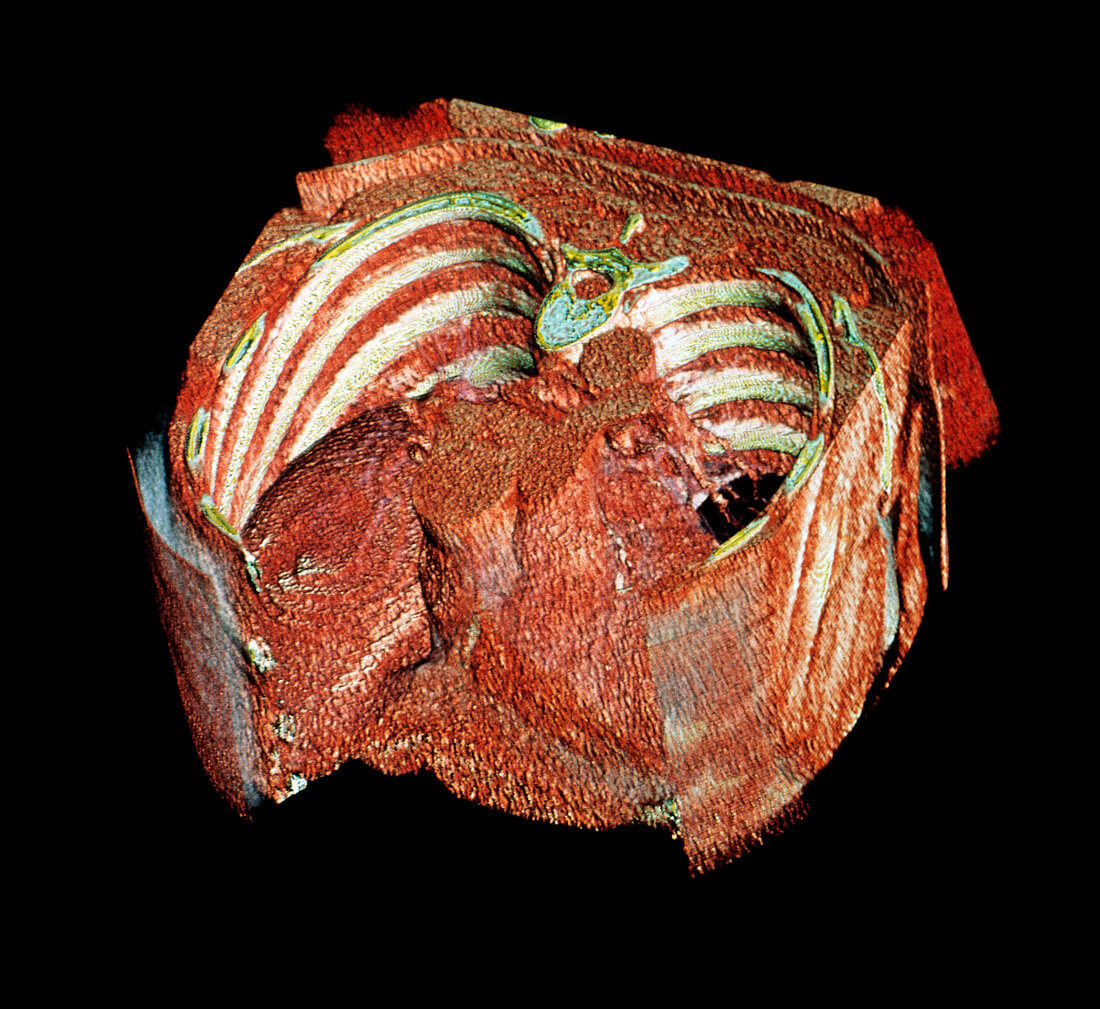
| Coronary artery disease. Coloured 3-D electron beam tomography (EBT) scan of a male heart (lower centre) showing calcium build-up in its arteries. EBT provides clearer and more detailed information about body tissues than CT (computed tomography) scans and reduces the time patients are exposed to dangerous radiation. EBT involves firing an electron beam against tungsten targets arranged in a 210 degree arc below the patient. These produce X-rays which pass through the patient. A computer is able to produce cross-sectional (slice) images of the body tissues,and 3-D images can be built up by combining many slices. The ribs (white) and intercostal muscles (red,between ribs) are seen | |
| Lizenzart: | Lizenzpflichtig |
| Credit: | Science Photo Library / King-Holmes, James |
| Bildgröße: | 3263 px × 2993 px |
| Modell-Rechte: | nicht erforderlich |
| Eigentums-Rechte: | nicht erforderlich |
| Restrictions: | - |
Preise für dieses Bild ab 15 €
Universitäten & Organisationen
(Informationsmaterial Digital, Informationsmaterial Print, Lehrmaterial Digital etc.)
ab 15 €
Redaktionell
(Bücher, Bücher: Sach- und Fachliteratur, Digitale Medien (redaktionell) etc.)
ab 30 €
Werbung
(Anzeigen, Aussenwerbung, Digitale Medien, Fernsehwerbung, Karten, Werbemittel, Zeitschriften etc.)
ab 55 €
Handelsprodukte
(bedruckte Textilie, Kalender, Postkarte, Grußkarte, Verpackung etc.)
ab 75 €
Pauschalpreise
Rechtepakete für die unbeschränkte Bildnutzung in Print oder Online
ab 495 €
Keywords
- 3-d,
- Anatomie,
- Bild,
- Bildgebung,
- Diagnose,
- Dreidimensional,
- EBCT,
- ebt,
- England,
- farbig,
- Gesundheitswesen,
- Herz,
- Hohlraum,
- Kalzium,
- London,
- Mann,
- Männlich,
- Medizin,
- medizinisch,
- menschlicher Körper,
- Muskel,
- Muskeln,
- Radiographie,
- Rippen,
- Röntgen,
- Scan,
- Technik,
- Technologie,
- technologisch,
- Thorax,
- Torso,
- Truhe,
- Wirbel,
- Wirbelsäule