LM cross-section of a Hurthle cell thyroid tumour
Bildnummer 11839568
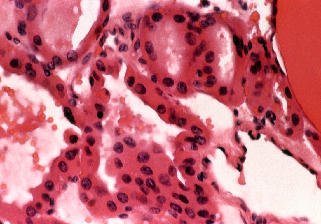
| Hurthle cell tumour. Light micrograph cross- section through a Hurthle cell tumour in a thyroid gland. The Hurthle cells are large and each has a large,dark nucleus. Hurthle cell tumours are be- nign but can be difficult to distinguish from cancerous tumours. Tumours are growths caused by the abnormal multiplication of cells. Benign tumours grow more slowly than cancers and do not spread to other parts of the body. However,the pressure exerted by the expanding tumour may damage surrounding tissue. Haemotoxylin and eosin stained sample. Magnification: x63 at 35mm size | |
| Lizenzart: | Lizenzpflichtig |
| Credit: | Science Photo Library / Ashton, Jonathan |
| Bildgröße: | 5024 px × 3513 px |
| Modell-Rechte: | nicht erforderlich |
| Eigentums-Rechte: | nicht erforderlich |
| Restrictions: | - |
Preise für dieses Bild ab 15 €
Universitäten & Organisationen
(Informationsmaterial Digital, Informationsmaterial Print, Lehrmaterial Digital etc.)
ab 15 €
Redaktionell
(Bücher, Bücher: Sach- und Fachliteratur, Digitale Medien (redaktionell) etc.)
ab 30 €
Werbung
(Anzeigen, Aussenwerbung, Digitale Medien, Fernsehwerbung, Karten, Werbemittel, Zeitschriften etc.)
ab 55 €
Handelsprodukte
(bedruckte Textilie, Kalender, Postkarte, Grußkarte, Verpackung etc.)
ab 75 €
Pauschalpreise
Rechtepakete für die unbeschränkte Bildnutzung in Print oder Online
ab 495 €