False-colour 3-D CT scan of kidney: hydronephrosis
Bildnummer 11839527
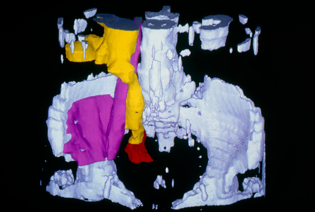
| False-colour three-dimensional computed tomography (CT) scan of the pelvic region,showing a kidney and ureter diseased with hydronephrosis. Bone is coloured white. The base of the right kidney is seen (top left,yellow) greatly distended with urine,due to an obstruction in the urinary tract,particularly the ureter. Here,the ureter has swelled along its tube length,to the point (red) at which it joins the bladder. The retaining of fluid caused by hydronephrosis can cause irrepair- able damage to the kidneys. In this image the pelvic psoas muscle (violet) is also seen,arising from thoracic vertebrae; it flexes the trunk | |
| Lizenzart: | Lizenzpflichtig |
| Credit: | Science Photo Library / CNRI |
| Bildgröße: | 4543 px × 3058 px |
| Modell-Rechte: | nicht erforderlich |
| Eigentums-Rechte: | nicht erforderlich |
| Restrictions: | - |
Preise für dieses Bild ab 15 €
Universitäten & Organisationen
(Informationsmaterial Digital, Informationsmaterial Print, Lehrmaterial Digital etc.)
ab 15 €
Redaktionell
(Bücher, Bücher: Sach- und Fachliteratur, Digitale Medien (redaktionell) etc.)
ab 30 €
Werbung
(Anzeigen, Aussenwerbung, Digitale Medien, Fernsehwerbung, Karten, Werbemittel, Zeitschriften etc.)
ab 55 €
Handelsprodukte
(bedruckte Textilie, Kalender, Postkarte, Grußkarte, Verpackung etc.)
ab 75 €
Pauschalpreise
Rechtepakete für die unbeschränkte Bildnutzung in Print oder Online
ab 495 €