Ophthalmoscopy of detached retina in patient's eye
Bildnummer 11838980
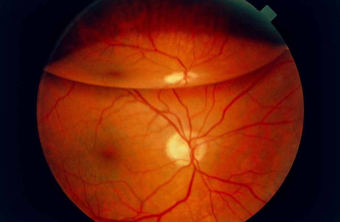
| Detached retina. Ophthalmoscope view of patient's eye with a detached retina. The retina has peeled away from it's choroid layer (seen at upper image) & formed a gas bubble. At lower centre,red blood vessels are seen emerging from the yellow optic disc. This is also where nerves enter the eye. To the left of the optic disc the dark macula is seen,where the largest concentration of light sensitive cells occur. Retinal detachment usually results from a tear in the retina,brought about by an injury or disease. This painless condition results in a "black drape" falling across the patient's vision. Surgical treatment is usually required to prevent loss of vision | |
| Lizenzart: | Lizenzpflichtig |
| Credit: | Science Photo Library / Ford, Sue |
| Bildgröße: | 4745 px × 3097 px |
| Modell-Rechte: | nicht erforderlich |
| Eigentums-Rechte: | nicht erforderlich |
| Restrictions: | - |
Preise für dieses Bild ab 15 €
Universitäten & Organisationen
(Informationsmaterial Digital, Informationsmaterial Print, Lehrmaterial Digital etc.)
ab 15 €
Redaktionell
(Bücher, Bücher: Sach- und Fachliteratur, Digitale Medien (redaktionell) etc.)
ab 30 €
Werbung
(Anzeigen, Aussenwerbung, Digitale Medien, Fernsehwerbung, Karten, Werbemittel, Zeitschriften etc.)
ab 55 €
Handelsprodukte
(bedruckte Textilie, Kalender, Postkarte, Grußkarte, Verpackung etc.)
ab 75 €
Pauschalpreise
Rechtepakete für die unbeschränkte Bildnutzung in Print oder Online
ab 495 €