Eczema
Bildnummer 11838764
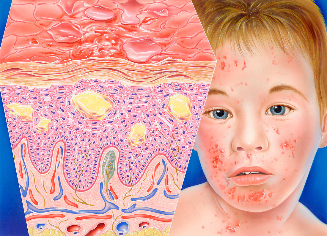
| Eczema. Artwork of a section through the epidermis of human skin affected by eczema. The boy's face shows the characteristic itchy pink rash on the skin's surface,accompanied by small,fluid- filled vesicles that weep and become crusted. Many factors can trigger the rash,but all tend to produce the same histological features. Fluid accumulates in the epidermis (folded pink layer),resulting in separation of the cells (spongiosis). Clumps of epidermal cells disintegrate to form vesicles (yellow). Blood vessels in the underlying dermis are dilated. The yellow layer at the bottom is subcutaneous fatty (adipose) tissue | |
| Lizenzart: | Lizenzpflichtig |
| Credit: | Science Photo Library / Bavosi, John |
| Bildgröße: | 4941 px × 3571 px |
| Modell-Rechte: | nicht erforderlich |
| Eigentums-Rechte: | nicht erforderlich |
| Restrictions: | - |
Preise für dieses Bild ab 15 €
Universitäten & Organisationen
(Informationsmaterial Digital, Informationsmaterial Print, Lehrmaterial Digital etc.)
ab 15 €
Redaktionell
(Bücher, Bücher: Sach- und Fachliteratur, Digitale Medien (redaktionell) etc.)
ab 30 €
Werbung
(Anzeigen, Aussenwerbung, Digitale Medien, Fernsehwerbung, Karten, Werbemittel, Zeitschriften etc.)
ab 55 €
Handelsprodukte
(bedruckte Textilie, Kalender, Postkarte, Grußkarte, Verpackung etc.)
ab 75 €
Pauschalpreise
Rechtepakete für die unbeschränkte Bildnutzung in Print oder Online
ab 495 €
Keywords
- atopisch,
- Ausschlag,
- Dermatologie,
- dermatologisch,
- Ekzem,
- entzündet,
- Entzündung,
- Epidermis,
- geduldig,
- Gesicht,
- Gesundheitswesen,
- Haut,
- Histologie,
- histologisch,
- Illustration,
- Jung,
- Junge,
- Kind,
- Kondition,
- Krankheit,
- Kunstwerk,
- Männlich,
- Medizin,
- medizinisch,
- Oberfläche,
- Schichten,
- Sektion,
- sektioniert,
- Spongiose,
- Störung,
- Vesikel