Ophthalmoscopy of maculopathy in diabetic's eye
Bildnummer 11838466
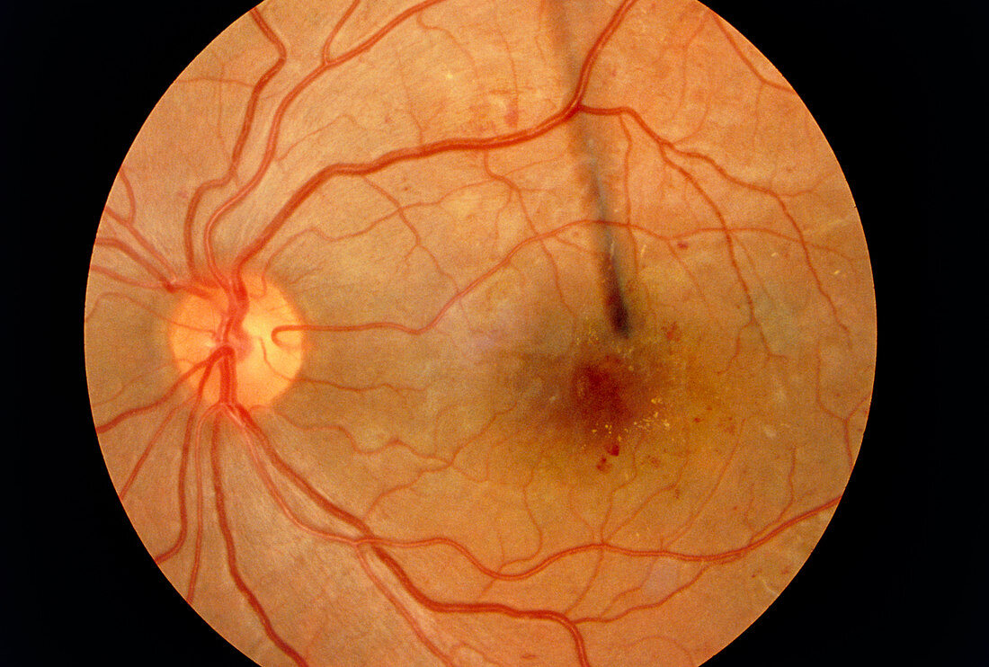
| Diabetic maculopathy. Ophthalmoscope view of the retina in the eye of a patient with diabetic maculopathy. A dark region at centre right is the macula,where the greatest concentration of light sensitive cells (rods & cones) are found. Other anatomical features seen include the optic disc,a yellow circle (at left) from which the nerves connecting the eye to the brain emerge. Red blood vessels are also seen emerging from the optic disc. In a normal retina the macula is seen as a pale yellow region. The diabetes in this patient is damaging the blood supply to small arteries and capillaries which supply the macula. This has lead to macular degeneration and vision loss | |
| Lizenzart: | Lizenzpflichtig |
| Credit: | Science Photo Library / Ford, Sue |
| Bildgröße: | 4962 px × 3345 px |
| Modell-Rechte: | nicht erforderlich |
| Eigentums-Rechte: | nicht erforderlich |
| Restrictions: | - |
Preise für dieses Bild ab 15 €
Universitäten & Organisationen
(Informationsmaterial Digital, Informationsmaterial Print, Lehrmaterial Digital etc.)
ab 15 €
Redaktionell
(Bücher, Bücher: Sach- und Fachliteratur, Digitale Medien (redaktionell) etc.)
ab 30 €
Werbung
(Anzeigen, Aussenwerbung, Digitale Medien, Fernsehwerbung, Karten, Werbemittel, Zeitschriften etc.)
ab 55 €
Handelsprodukte
(bedruckte Textilie, Kalender, Postkarte, Grußkarte, Verpackung etc.)
ab 75 €
Pauschalpreise
Rechtepakete für die unbeschränkte Bildnutzung in Print oder Online
ab 495 €