Brain arteriovenous malformation,MRA
Bildnummer 11838349
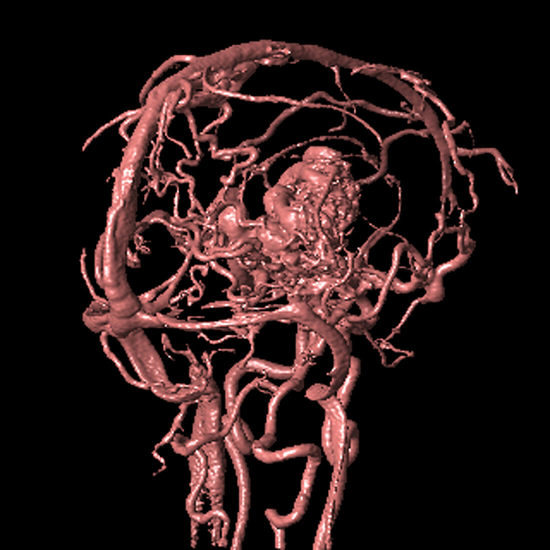
| Brain arteriovenous malformation. Image 3 of 6. Coloured magnetic resonance angiography (MRA) scan of the brain of a 31 year old man (side view). The rear of the head is at left. The image shows a right thalamic AVM as a knotted red mass (centre). An AVM is a congenital malformation of the blood vessels (arteries and veins) that can rupture and bleed inside of the head and cause seizures,headaches,stroke-like symptoms,and other fatal complications. MRA is a non-invasive technique to image blood vessels in the body that uses a combination of a very strong magnetic field and radiofrequency (RF) pulses to image the flowing blood. For all images in this rotational sequence,see M136/300 - M136/305 | |
| Lizenzart: | Lizenzpflichtig |
| Credit: | Science Photo Library / Fraser, Simon |
| Bildgröße: | 2647 px × 2647 px |
| Modell-Rechte: | nicht erforderlich |
| Eigentums-Rechte: | nicht erforderlich |
| Restrictions: | - |
Preise für dieses Bild ab 15 €
Universitäten & Organisationen
(Informationsmaterial Digital, Informationsmaterial Print, Lehrmaterial Digital etc.)
ab 15 €
Redaktionell
(Bücher, Bücher: Sach- und Fachliteratur, Digitale Medien (redaktionell) etc.)
ab 30 €
Werbung
(Anzeigen, Aussenwerbung, Digitale Medien, Fernsehwerbung, Karten, Werbemittel, Zeitschriften etc.)
ab 55 €
Handelsprodukte
(bedruckte Textilie, Kalender, Postkarte, Grußkarte, Verpackung etc.)
ab 75 €
Pauschalpreise
Rechtepakete für die unbeschränkte Bildnutzung in Print oder Online
ab 495 €
Keywords
- 30er Jahre,
- abnormal,
- angeboren,
- Angiografie,
- Angiogramm,
- Arterien,
- arteriovenöse Malformation,
- AVM,
- Bild,
- Blut,
- Blutgefäß,
- Blutgefäße,
- cerebral,
- Diagnose,
- dreißiger Jahre,
- Erwachsene,
- Gefäß,
- Gehirn,
- gutartig,
- Knoten,
- Kondition,
- Krankheit,
- Kreislauf,
- Magnetresonanzangiographie,
- Magnetresonanztomografie,
- Mann,
- Männlich,
- Medizin,
- medizinisch,
- menschlicher Körper,
- MRA,
- MRI,
- MRT-Untersuchung,
- Neuroimaging,
- Reihenfolge,
- Scanner,
- Seitenansicht,
- Störung,
- Thalamus,
- Tumor,
- ungesund,
- vaskulär,
- Vene,
- Venen