Brain arteriovenous malformation,MRI
Bildnummer 11838345
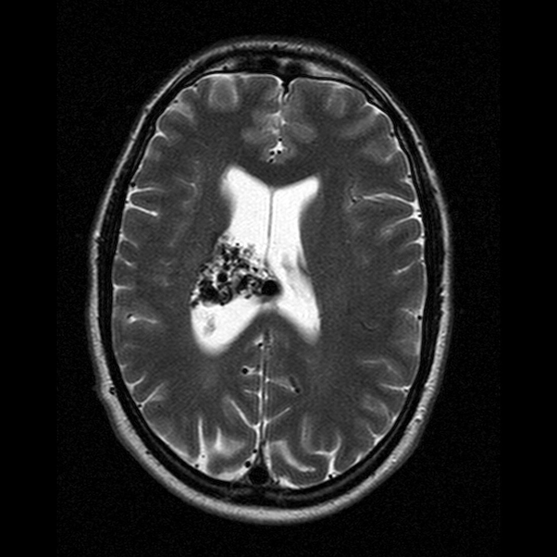
| Brain arteriovenous malformation (AVM). Magnetic resonance imaging (MRI) scan of an axial (horizontal) section through the brain of a 31 year old man. The image shows a right thalamic AVM as a dark grey mass on a white background (centre). The black areas within it indicate the damage caused by the AVM; they are flow voids,areas that no longer receive blood flow. An AVM is a congenital malformation of the blood vessels (arteries and veins) that can rupture and bleed inside of the head and cause seizures,headaches,stroke-like symptoms,and other fatal complications. MRI uses radio waves and a magnet to obtain "slice" body images | |
| Lizenzart: | Lizenzpflichtig |
| Credit: | Science Photo Library / Fraser, Simon |
| Bildgröße: | 2647 px × 2647 px |
| Modell-Rechte: | nicht erforderlich |
| Eigentums-Rechte: | nicht erforderlich |
| Restrictions: | - |
Preise für dieses Bild ab 15 €
Universitäten & Organisationen
(Informationsmaterial Digital, Informationsmaterial Print, Lehrmaterial Digital etc.)
ab 15 €
Redaktionell
(Bücher, Bücher: Sach- und Fachliteratur, Digitale Medien (redaktionell) etc.)
ab 30 €
Werbung
(Anzeigen, Aussenwerbung, Digitale Medien, Fernsehwerbung, Karten, Werbemittel, Zeitschriften etc.)
ab 55 €
Handelsprodukte
(bedruckte Textilie, Kalender, Postkarte, Grußkarte, Verpackung etc.)
ab 75 €
Pauschalpreise
Rechtepakete für die unbeschränkte Bildnutzung in Print oder Online
ab 495 €
Keywords
- 30er Jahre,
- abnormal,
- angeboren,
- Arterien,
- arteriovenöse Malformation,
- AVM,
- axial,
- Blut,
- Blutgefäß,
- Blutgefäße,
- cerebral,
- Diagnose,
- dreißiger Jahre,
- Einfarbig,
- Erwachsene,
- Gefäß,
- Gehirn,
- gutartig,
- horizontal,
- Knoten,
- Kondition,
- Krankheit,
- Kreislauf,
- Magnetresonanztomografie,
- Mann,
- Männlich,
- Medizin,
- medizinisch,
- menschlicher Körper,
- MRI,
- MRT-Untersuchung,
- Neuroimaging,
- Organ,
- Querschnitt,
- Scanner,
- Schwarz und weiß,
- Sektion,
- Störung,
- Thalamus,
- Tumor,
- ungesund,
- vaskulär,
- Vene,
- Venen