Vertebral haemangioma,MRI scan
Bildnummer 11838321
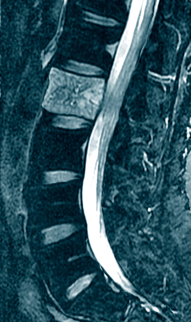
| Vertebral haemangioma. Coloured sagittal magnetic resonance imaging (MRI) scan of the lumbar spine of a 56-year-old patient with a vertebral haemangioma. The front of the spine is at left in this view from the side. The spinal cord (white) is seen to the right of the vertebral blocks (square shapes) that form the spinal column. A haemangioma is a benign blood vessel tumour,here seen at upper left inside one of the spinal bones (vertebrae). The other spinal vertebrae (around 6 seen here) are dark,but the one at upper left (the L2 vertebra) is white due to the presence of the haemangioma,which is causing pain by compressing the spinal cord. In such cases,the haemangioma is surgically removed | |
| Lizenzart: | Lizenzpflichtig |
| Credit: | Science Photo Library / Zephyr |
| Bildgröße: | 2340 px × 3933 px |
| Modell-Rechte: | nicht erforderlich |
| Eigentums-Rechte: | nicht erforderlich |
| Restrictions: | - |
Preise für dieses Bild ab 15 €
Universitäten & Organisationen
(Informationsmaterial Digital, Informationsmaterial Print, Lehrmaterial Digital etc.)
ab 15 €
Redaktionell
(Bücher, Bücher: Sach- und Fachliteratur, Digitale Medien (redaktionell) etc.)
ab 30 €
Werbung
(Anzeigen, Aussenwerbung, Digitale Medien, Fernsehwerbung, Karten, Werbemittel, Zeitschriften etc.)
ab 55 €
Handelsprodukte
(bedruckte Textilie, Kalender, Postkarte, Grußkarte, Verpackung etc.)
ab 75 €
Pauschalpreise
Rechtepakete für die unbeschränkte Bildnutzung in Print oder Online
ab 495 €
Keywords
- 50er Jahre,
- Blau,
- Diagnose,
- Einfarbig,
- Erwachsene,
- farbig,
- Fünfziger Jahre,
- geduldig,
- gefärbt,
- Gesundheitswesen,
- Knochen,
- Kondition,
- Krankheit,
- L2,
- Lendenwirbelsäule,
- Magnetresonanztomografie,
- Medizin,
- medizinisch,
- menschlicher Körper,
- mittleren Alters,
- MRT-Untersuchung,
- Osteologie,
- Rücken,
- Rückgrat,
- Scanner,
- Seite,
- Skelett-,
- Störung,
- Tumor,
- unterer Rücken,
- vaskulär,
- vertebral,
- Wachstum,
- Wirbel,
- Wirbelsäule,
- Wirbelsäulen-