Stroke,CT scans
Bildnummer 11838295
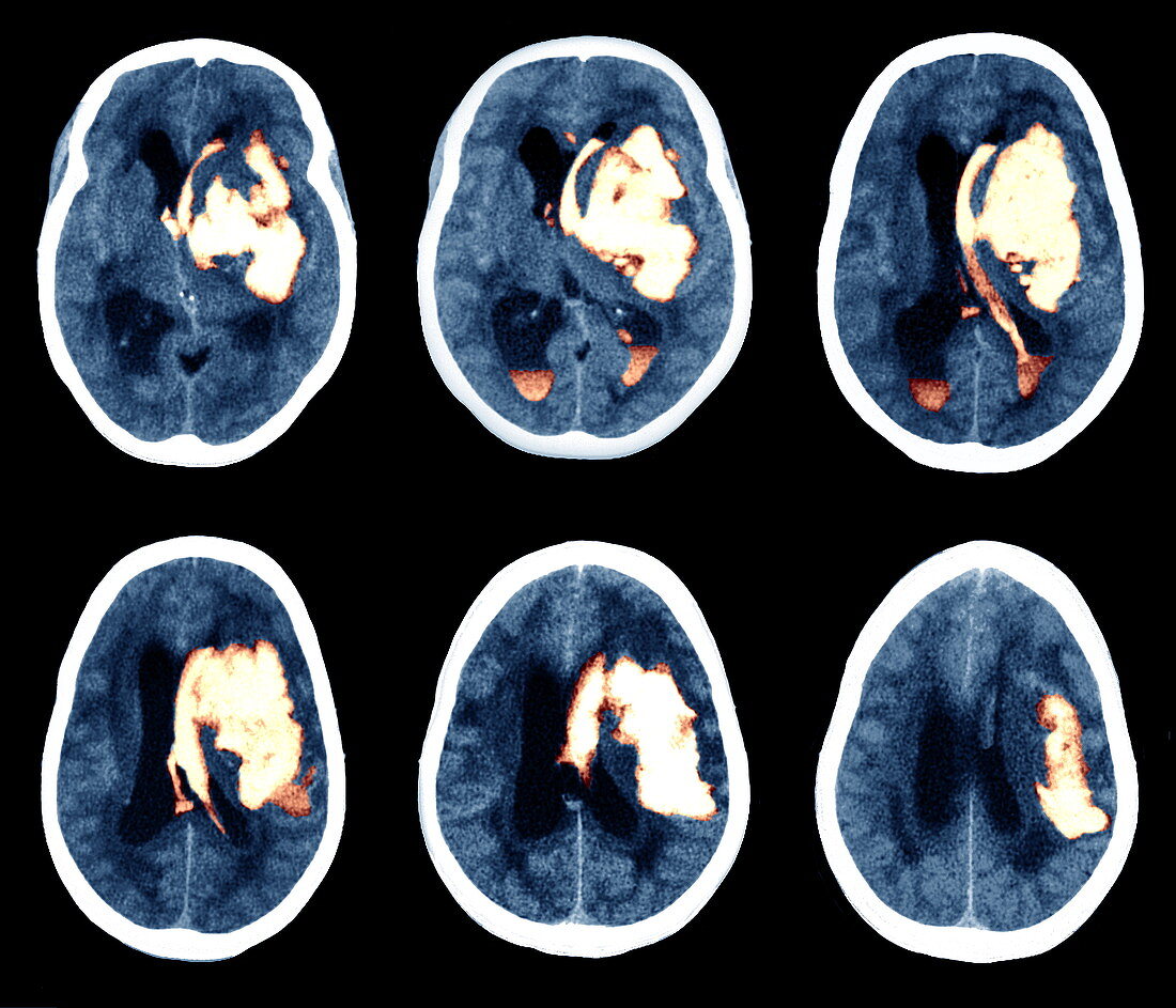
| Stroke. Coloured computed tomography (CT) scans of axial (horizontal) sections through different levels of a stroke victim's brain. The stroke has resulted in internal bleeding (white/orange). The front of the brain is at top in each image. The mass of blood (haematoma) extends up and down in the brain as well as across the left hemisphere,and has ruptured the ventricles (black) that carry the brain's cerebrospinal fluid. This stroke occurred 15 days after drug treatment to prevent a stroke was suspended. Brain damage: aphasia (loss of speech) and hemiplegia (paralysis of one side of the body) occurred. Common causes of a stroke are high blood pressure and arterial disease | |
| Lizenzart: | Lizenzpflichtig |
| Credit: | Science Photo Library / Zephyr |
| Bildgröße: | 4134 px × 3543 px |
| Modell-Rechte: | nicht erforderlich |
| Eigentums-Rechte: | nicht erforderlich |
| Restrictions: | - |
Preise für dieses Bild ab 15 €
Universitäten & Organisationen
(Informationsmaterial Digital, Informationsmaterial Print, Lehrmaterial Digital etc.)
ab 15 €
Redaktionell
(Bücher, Bücher: Sach- und Fachliteratur, Digitale Medien (redaktionell) etc.)
ab 30 €
Werbung
(Anzeigen, Aussenwerbung, Digitale Medien, Fernsehwerbung, Karten, Werbemittel, Zeitschriften etc.)
ab 55 €
Handelsprodukte
(bedruckte Textilie, Kalender, Postkarte, Grußkarte, Verpackung etc.)
ab 75 €
Pauschalpreise
Rechtepakete für die unbeschränkte Bildnutzung in Print oder Online
ab 495 €
Keywords
- axial,
- Beschädigt,
- Blut,
- Blutgefäß,
- Blutung,
- cerebral,
- Computertomographie,
- ct,
- CVA,
- Diagnose,
- Ebenen,
- farbig,
- geduldig,
- Gesundheitswesen,
- Hämatom,
- Hemiplegie,
- Hemisphären,
- Kleinhirn,
- Kondition,
- Krankheit,
- Kreislauf,
- Lähmung,
- Medizin,
- medizinisch,
- Neuroimaging,
- Neurologie,
- Reihenfolge,
- Scan,
- Schlaganfall,
- Sektion,
- sektioniert,
- Serie,
- Störung,
- vaskulär,
- Ventrikel,
- zentrales Nervensystem