Stroke,CT scan
Bildnummer 11838294
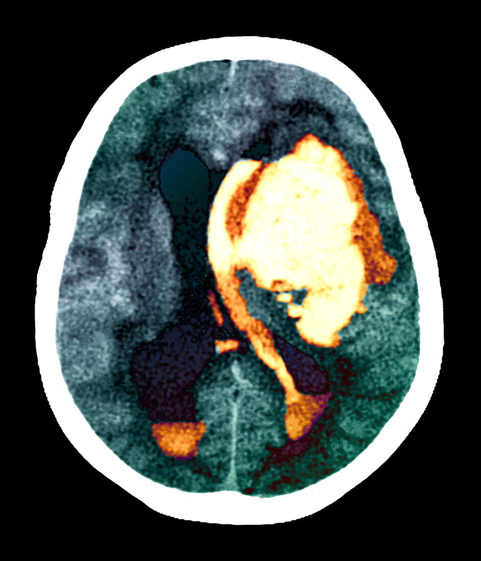
| Stroke. Coloured computed tomography (CT) scan of an axial (horizontal) section through a patient's brain showing internal bleeding (yellow/orange) due to a cerebrovascular accident (CVA,stroke). The front of the brain is at top. The main mass of blood (haematoma) is at upper right. Left of this,blood abnormally fills a ruptured ventricle,and left of that,a normal ventricle (black) helps to circulate the brain's cerebrospinal fluid. This stroke occurred 15 days after drug treatment to prevent a stroke was halted. Aphasia (loss of speech) and hemiplegia (paralysis of one side of the body) occurred. Common causes of a stroke are high blood pressure and arterial disease | |
| Lizenzart: | Lizenzpflichtig |
| Credit: | Science Photo Library / Zephyr |
| Bildgröße: | 3543 px × 4134 px |
| Modell-Rechte: | nicht erforderlich |
| Eigentums-Rechte: | nicht erforderlich |
| Restrictions: | - |
Preise für dieses Bild ab 15 €
Universitäten & Organisationen
(Informationsmaterial Digital, Informationsmaterial Print, Lehrmaterial Digital etc.)
ab 15 €
Redaktionell
(Bücher, Bücher: Sach- und Fachliteratur, Digitale Medien (redaktionell) etc.)
ab 30 €
Werbung
(Anzeigen, Aussenwerbung, Digitale Medien, Fernsehwerbung, Karten, Werbemittel, Zeitschriften etc.)
ab 55 €
Handelsprodukte
(bedruckte Textilie, Kalender, Postkarte, Grußkarte, Verpackung etc.)
ab 75 €
Pauschalpreise
Rechtepakete für die unbeschränkte Bildnutzung in Print oder Online
ab 495 €
Keywords
- axial,
- Beschädigt,
- Blut,
- Blutgefäß,
- Blutung,
- cerebral,
- Computertomographie,
- ct,
- CVA,
- Diagnose,
- farbig,
- Gesundheitswesen,
- Hämatom,
- Hemiplegie,
- Hemisphären,
- Kleinhirn,
- Kondition,
- Krankheit,
- Kreislauf,
- Lähmung,
- Medizin,
- medizinisch,
- Neuroimaging,
- Neurologie,
- Scan,
- Schlaganfall,
- Sektion,
- sektioniert,
- Störung,
- vaskulär,
- Ventrikel,
- zentrales Nervensystem