Blood vessel tumour
Bildnummer 11838283
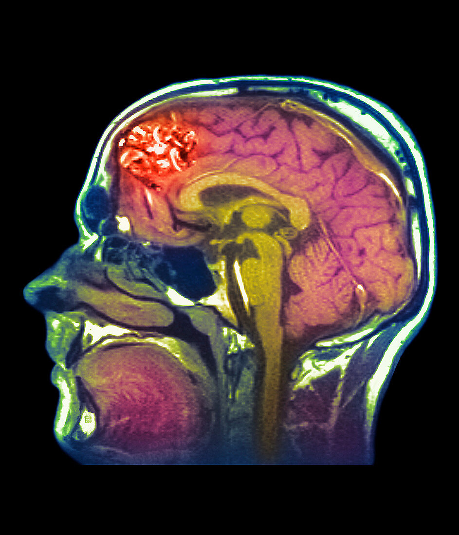
| Blood vessel tumour. Coloured magnetic resonance imaging (MRI) head scan showing an arteriovenous malformation (AVM,red,upper left). The AVM is in the brain (pink & yellow) of a 20-year-old patient in this sagittal (vertical) image. This benign (non-cancerous) tumour,also known as an arteriovenous angioma,is formed from a clump of distended blood vessels. It may compress the brain,resulting in epilepsy,or cause a haemorrhage if a vessel bursts. AVMs are surgically removed or destroyed by radiotherapy. MRI scans produce slice images through tissues using a powerful magnet and pulses of radio waves | |
| Lizenzart: | Lizenzpflichtig |
| Credit: | Science Photo Library / RNC, NEWCASTLE / SIMON FRASER |
| Bildgröße: | 3000 px × 3500 px |
| Modell-Rechte: | nicht erforderlich |
| Eigentums-Rechte: | nicht erforderlich |
| Restrictions: | - |
Preise für dieses Bild ab 15 €
Universitäten & Organisationen
(Informationsmaterial Digital, Informationsmaterial Print, Lehrmaterial Digital etc.)
ab 15 €
Redaktionell
(Bücher, Bücher: Sach- und Fachliteratur, Digitale Medien (redaktionell) etc.)
ab 30 €
Werbung
(Anzeigen, Aussenwerbung, Digitale Medien, Fernsehwerbung, Karten, Werbemittel, Zeitschriften etc.)
ab 55 €
Handelsprodukte
(bedruckte Textilie, Kalender, Postkarte, Grußkarte, Verpackung etc.)
ab 75 €
Pauschalpreise
Rechtepakete für die unbeschränkte Bildnutzung in Print oder Online
ab 495 €