Colour PET brain scans of stroke vs normal patient
Bildnummer 11838243
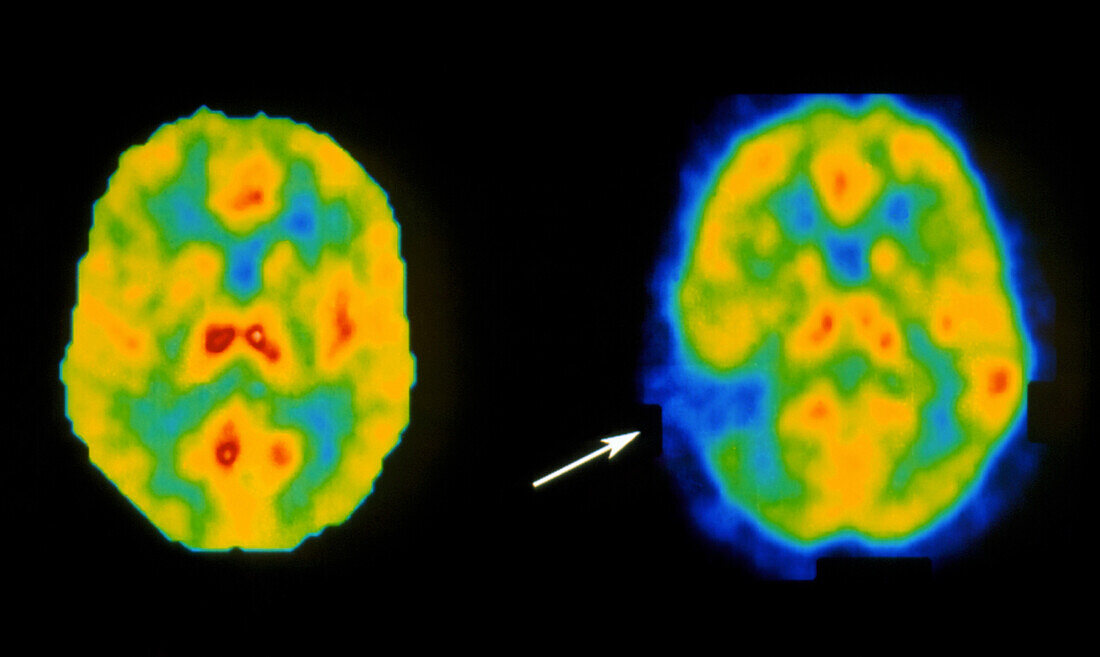
| Stroke with speech loss. Colour Positron Emission Tomography (PET) brain scans of a healthy patient (at left) versus stroke patient. Horizontal slices are seen through the temporal lobes. The stroke affected brain shows a lesion (arrowed),an area of brain damage with low blood flow and activity. This affected area has led to "aphasia",partial loss of speech. Colour-coding is: high brain activity (red/yellow); low activity (blue). Stroke or cerebrovascular accident (CVA),is a rupture or blockage of a blood vessel leading to brain damage and sometimes paralysis. PET scanning shows blood flow and metabolic activity in the brain | |
| Lizenzart: | Lizenzpflichtig |
| Credit: | Science Photo Library / Wellcome Dept. of Cognitive Neurology |
| Bildgröße: | 4152 px × 2480 px |
| Modell-Rechte: | nicht erforderlich |
| Eigentums-Rechte: | nicht erforderlich |
| Restrictions: | - |
Preise für dieses Bild ab 15 €
Universitäten & Organisationen
(Informationsmaterial Digital, Informationsmaterial Print, Lehrmaterial Digital etc.)
ab 15 €
Redaktionell
(Bücher, Bücher: Sach- und Fachliteratur, Digitale Medien (redaktionell) etc.)
ab 30 €
Werbung
(Anzeigen, Aussenwerbung, Digitale Medien, Fernsehwerbung, Karten, Werbemittel, Zeitschriften etc.)
ab 55 €
Handelsprodukte
(bedruckte Textilie, Kalender, Postkarte, Grußkarte, Verpackung etc.)
ab 75 €
Pauschalpreise
Rechtepakete für die unbeschränkte Bildnutzung in Print oder Online
ab 495 €