X-ray tomography scan of brain showing haemorrhage
Bildnummer 11838220
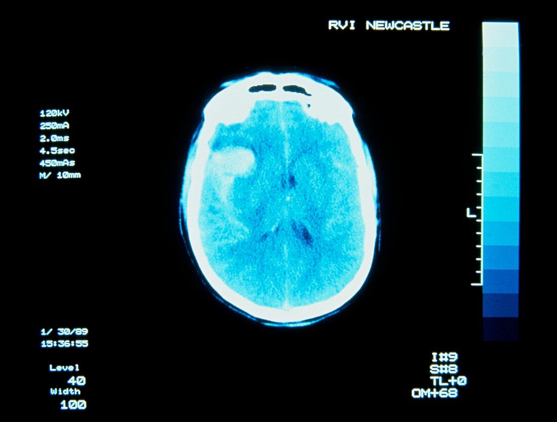
| Computed X-ray tomography (CT) scan of the brain showing a subarachnoid haemorrhage,the result of a ruptured blood vessel & one cause of stroke. The haemorrhage appears as the light area over the right side of the brain (left on image). In CT,a narrow X-ray beam is directed through the subject towards a diametrically-opposed detector. A series of "slices" is made,with source & detector moving synchronously around the subject. Measurements of transmitted X-rays are processed by computer to reveal how elements of tissue in each "slice" attenuate X-rays - the basis for constructing the image. Courtesy of Radiography Dept.,Royal Victoria Infirmary,Newcastle-upon-Tyne | |
| Lizenzart: | Lizenzpflichtig |
| Credit: | Science Photo Library / Fraser, Simon |
| Bildgröße: | 4843 px × 3670 px |
| Modell-Rechte: | nicht erforderlich |
| Eigentums-Rechte: | nicht erforderlich |
| Restrictions: | - |
Preise für dieses Bild ab 15 €
Universitäten & Organisationen
(Informationsmaterial Digital, Informationsmaterial Print, Lehrmaterial Digital etc.)
ab 15 €
Redaktionell
(Bücher, Bücher: Sach- und Fachliteratur, Digitale Medien (redaktionell) etc.)
ab 30 €
Werbung
(Anzeigen, Aussenwerbung, Digitale Medien, Fernsehwerbung, Karten, Werbemittel, Zeitschriften etc.)
ab 55 €
Handelsprodukte
(bedruckte Textilie, Kalender, Postkarte, Grußkarte, Verpackung etc.)
ab 75 €
Pauschalpreise
Rechtepakete für die unbeschränkte Bildnutzung in Print oder Online
ab 495 €