Bone cancer,neck MRI scan
Bildnummer 11837978
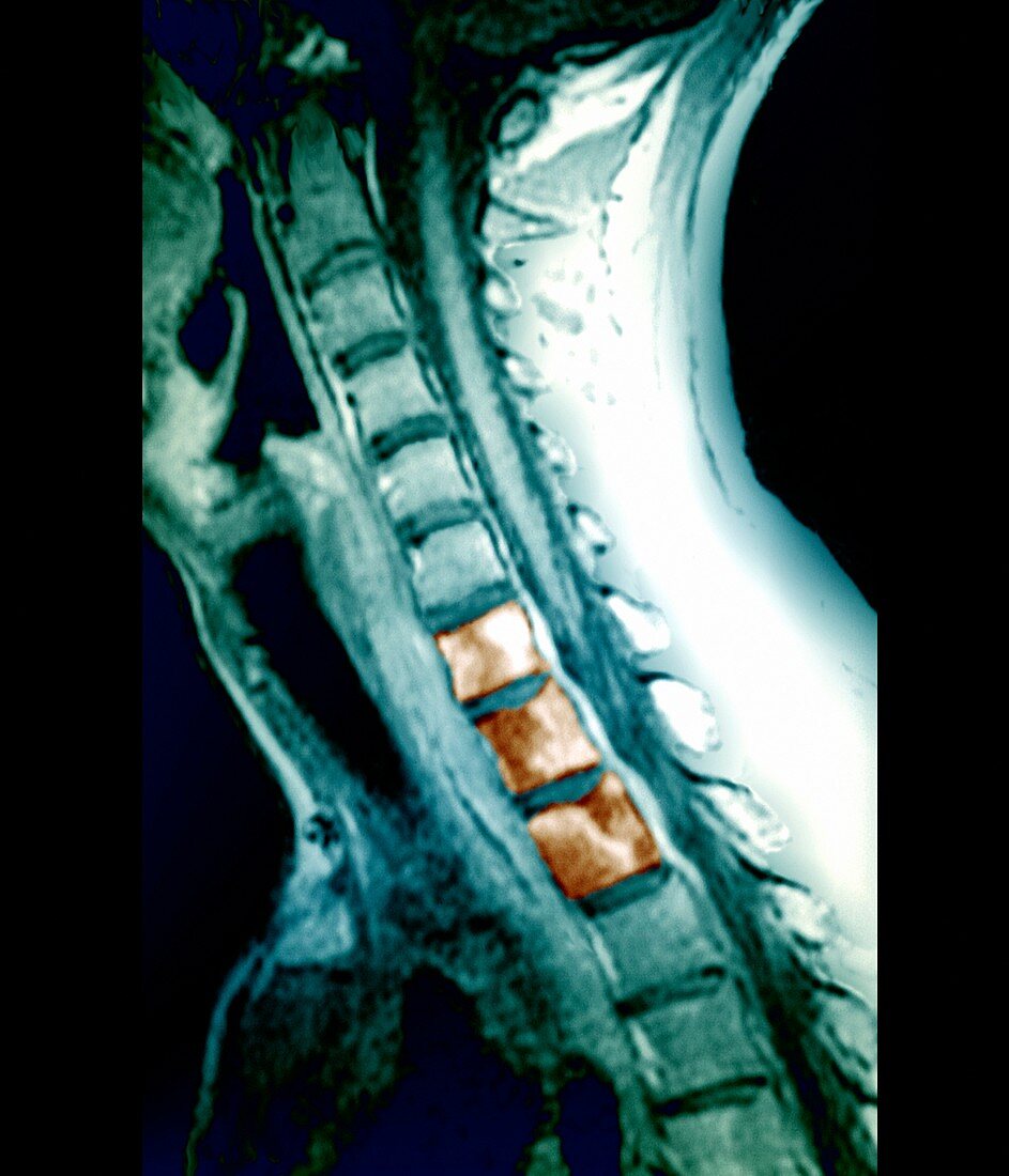
| Bone cancer. Coloured sagittal (side) magnetic resonance imaging (MRI) scan through the neck of a patient with secondary bone cancer of three spinal vertebrae (orange). The front of the body is at left. The cancerous vertebrae are (from bottom): the top two thoracic (chest) vertebrae (T2 and T1) and the seventh cervical (neck) vertebra (C7). The spinal cord is seen in the space at right of the highlighted portions of the vertebrae. The cancer has spread (metastasized) from a site of primary cancer in the lung. Spread of cancer to the spine is common. The prognosis is very poor. Treatments such as steroid drugs and radiotherapy are aimed at preserving function and quality of life | |
| Lizenzart: | Lizenzpflichtig |
| Credit: | Science Photo Library / Zephyr |
| Bildgröße: | 3543 px × 4131 px |
| Modell-Rechte: | nicht erforderlich |
| Eigentums-Rechte: | nicht erforderlich |
| Restrictions: | - |
Preise für dieses Bild ab 15 €
Universitäten & Organisationen
(Informationsmaterial Digital, Informationsmaterial Print, Lehrmaterial Digital etc.)
ab 15 €
Redaktionell
(Bücher, Bücher: Sach- und Fachliteratur, Digitale Medien (redaktionell) etc.)
ab 30 €
Werbung
(Anzeigen, Aussenwerbung, Digitale Medien, Fernsehwerbung, Karten, Werbemittel, Zeitschriften etc.)
ab 55 €
Handelsprodukte
(bedruckte Textilie, Kalender, Postkarte, Grußkarte, Verpackung etc.)
ab 75 €
Pauschalpreise
Rechtepakete für die unbeschränkte Bildnutzung in Print oder Online
ab 495 €
Keywords
- Bild,
- Diagnose,
- dorsal,
- farbig,
- Gebärmutterhals-,
- geduldig,
- Gesundheitswesen,
- Hals,
- Kondition,
- Krankheit,
- krebsartig,
- Lunge,
- Magnetresonanztomografie,
- maligne,
- Malignom,
- Medizin,
- medizinisch,
- Metastase,
- MRT-Untersuchung,
- Osteologie,
- osteologisch,
- Rücken,
- Rückgrat,
- Seite,
- Sektion,
- sektioniert,
- sekundär,
- Störung,
- Tumor,
- Wachstum,
- Wirbel,
- Wirbelsäule,
- Wirbelsäulen-