Liver cancer,Doppler ultrasound
Bildnummer 11837938
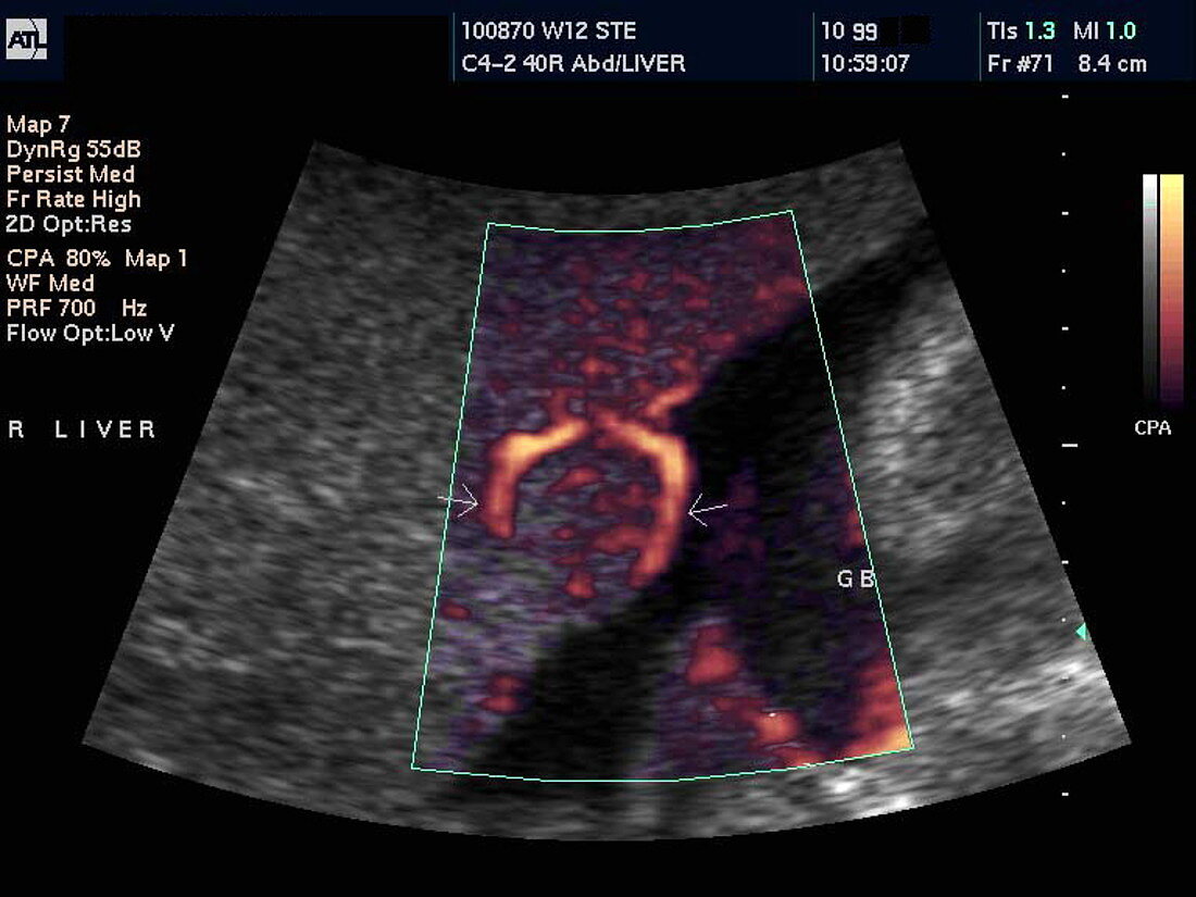
| Liver cancer. Power Doppler ultrasound scan of increased blood flow (orange) to a cancer in a 29- year-old woman's liver. Part of the liver is seen as the large grey area at left and top. The cancer is about 2 centimetres across (centre),shown by the arrows and its blood supply. The black area is the gall bladder. This liver cancer (hepatoma) resulted from cirrhosis,a condition caused by damage such as that from alcoholism and hepatitis. Doppler ultrasound scanning detects moving fluids,such as blood,by the frequency change they cause when reflecting high-frequency sound waves. Power Doppler scans use colour to show the moving fluid,but do not show the direction of the flow | |
| Lizenzart: | Lizenzpflichtig |
| Credit: | Science Photo Library / Fraser, Simon |
| Bildgröße: | 768 px × 576 px |
| Modell-Rechte: | nicht erforderlich |
| Eigentums-Rechte: | nicht erforderlich |
| Restrictions: | - |
Preise für dieses Bild ab 15 €
Universitäten & Organisationen
(Informationsmaterial Digital, Informationsmaterial Print, Lehrmaterial Digital etc.)
ab 15 €
Redaktionell
(Bücher, Bücher: Sach- und Fachliteratur, Digitale Medien (redaktionell) etc.)
ab 30 €
Werbung
(Anzeigen, Aussenwerbung, Digitale Medien, Fernsehwerbung, Karten, Werbemittel, Zeitschriften etc.)
ab 55 €
Handelsprodukte
(bedruckte Textilie, Kalender, Postkarte, Grußkarte, Verpackung etc.)
ab 75 €
Pauschalpreise
Rechtepakete für die unbeschränkte Bildnutzung in Print oder Online
ab 495 €
Keywords
- Abdomen,
- alkoholisch,
- Alkoholismus,
- Bauch,
- Bild,
- Blutfluss,
- Diagnose,
- Farbe,
- Frau,
- Galle,
- geduldig,
- Gefäß,
- Gefäße,
- Gefäßsystem,
- Gesundheitswesen,
- Hepatom,
- Kondition,
- Krankheit,
- krebsartig,
- Kreislauf,
- Leber,
- Leistung,
- maligne,
- Malignom,
- Medizin,
- medizinisch,
- Organ,
- primär,
- Scan,
- Scannen,
- Sonographie,
- Störung,
- Tumor,
- Ultraschall,
- Wachstum,
- Weiblich,
- Zirrhose