Spinal cord cancer MRI
Bildnummer 11837909
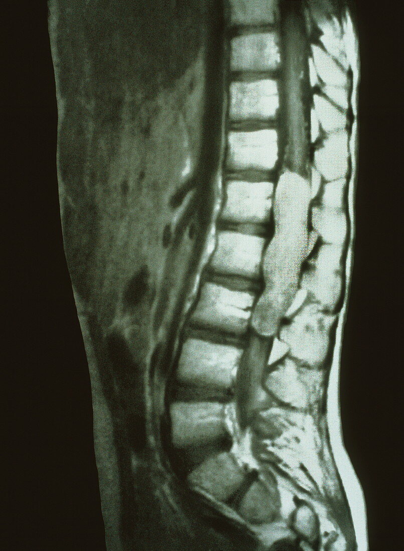
| Spinal cord cancer. Magnetic resonance imaging (MRI) scan showing a cancerous tumour growing in the spinal cord of a 10-year-old girl. This is a sagittal view,a vertical slice image seen from the side. The malignant tumour (pale vertical oblong,centre right) lies next to the three white blocks,parts of the vertebrae of the spinal column (down centre). The spinal cord runs up through the middle of the vertebrae,which are seen on either side of the tumour. MRI scans produce slice images of the body using pulses of radio waves and a powerful magnet | |
| Lizenzart: | Lizenzpflichtig |
| Credit: | Science Photo Library / Kulyk, Mehau |
| Bildgröße: | 3215 px × 4385 px |
| Modell-Rechte: | nicht erforderlich |
| Eigentums-Rechte: | nicht erforderlich |
| Restrictions: | - |
Preise für dieses Bild ab 15 €
Universitäten & Organisationen
(Informationsmaterial Digital, Informationsmaterial Print, Lehrmaterial Digital etc.)
ab 15 €
Redaktionell
(Bücher, Bücher: Sach- und Fachliteratur, Digitale Medien (redaktionell) etc.)
ab 30 €
Werbung
(Anzeigen, Aussenwerbung, Digitale Medien, Fernsehwerbung, Karten, Werbemittel, Zeitschriften etc.)
ab 55 €
Handelsprodukte
(bedruckte Textilie, Kalender, Postkarte, Grußkarte, Verpackung etc.)
ab 75 €
Pauschalpreise
Rechtepakete für die unbeschränkte Bildnutzung in Print oder Online
ab 495 €
Keywords
- Anatomie,
- Bild,
- Diagnose,
- Erkrankung,
- Gesundheitswesen,
- Kind,
- Kondition,
- Krankheit,
- krebsartig,
- Mädchen,
- Magnetresonanztomografie,
- maligne,
- Malignom,
- Medizin,
- medizinisch,
- MRT-Untersuchung,
- Nerv,
- Nervensystem,
- Rückenmark,
- Rückgrat,
- Schwarz und weiß,
- Seitenansicht,
- Störung,
- Tumor,
- vertebral,
- Weiblich,
- Wirbelsäule