Coloured MRI brain scan showing an area of cancer
Bildnummer 11837864
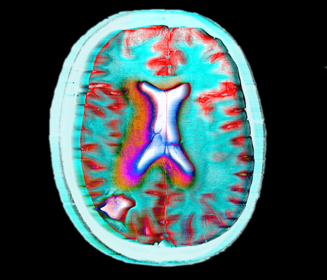
| Brain cancer. Coloured magnetic resonance imaging (MRI) scan of an axial section through a brain with a cancerous tumour (white,lower left). This malignant area of abnormal growth may be putting pressure on surrounding brain tissue,resulting in sensory disturbances,headaches,nausea and epileptic fits. The brain's ventricles (white),its fluid-filled cavities,are at centre. Brain tumours are surgically removed unless they are inaccessible,in which case radiotherapy and chemotherapy are used. MRI scanners produce slice images of the body using radio signals and a magnetic field. The scans are useful in revealing brain tumours and in planning brain surgery | |
| Lizenzart: | Lizenzpflichtig |
| Credit: | Science Photo Library / Gustoimages |
| Bildgröße: | 2800 px × 2400 px |
| Modell-Rechte: | nicht erforderlich |
| Eigentums-Rechte: | nicht erforderlich |
| Restrictions: | - |
Preise für dieses Bild ab 15 €
Universitäten & Organisationen
(Informationsmaterial Digital, Informationsmaterial Print, Lehrmaterial Digital etc.)
ab 15 €
Redaktionell
(Bücher, Bücher: Sach- und Fachliteratur, Digitale Medien (redaktionell) etc.)
ab 30 €
Werbung
(Anzeigen, Aussenwerbung, Digitale Medien, Fernsehwerbung, Karten, Werbemittel, Zeitschriften etc.)
ab 55 €
Handelsprodukte
(bedruckte Textilie, Kalender, Postkarte, Grußkarte, Verpackung etc.)
ab 75 €
Pauschalpreise
Rechtepakete für die unbeschränkte Bildnutzung in Print oder Online
ab 495 €